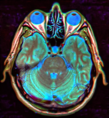
Empty sella syndrome is the condition when the pituitary gland shrinks or becomes flattened, filling the sella turcica with cerebrospinal fluid instead of the normal pituitary.[2] It can be discovered as part of the diagnostic workup of pituitary disorders, or as an incidental finding when imaging the brain.[1]
| Empty sella syndrome | |
|---|---|
| Other names | Pituitary - empty sella syndrome[1] |
 | |
| MRI of Empty Sella | |
| Specialty | Endocrinology |
| Symptoms | Cryptorchidism |
| Causes | Arachnoid presses down on gland (another possibility is a Tumor, Radiation therapy)[1] |
| Diagnostic method | MRI, CT scan[1] |
| Medication | Manage abnormal hormone levels[1] |
Signs and symptoms
editIf there are symptoms, people with empty sella syndrome can have headaches and vision loss. Additional symptoms would be associated with hypopituitarism.[3][4] Additional symptoms are as follows:[citation needed]
- Abnormality of the middle ear ossicles
- Cryptorchidism
- Dolichocephaly
- Arnold-Chiari type I malformation
- Meningocele
- Patent ductus arteriosus
- Muscular hypotonia
- Platybasia
Cause
editThe cause of this condition is divided into primary and secondary, as follows:
- The cause of this condition in terms of secondary empty sella syndrome happens when a tumor or surgery damages the gland, this is an acquired manner of the condition.[1]
- patients with idiopathic intracranial hypertension will have empty sella on MRI[5]
- The cause of primary empty sella syndrome is a congenital defect (diaphragma sellae)[6]
Mechanism
editThe normal mechanism of the pituitary gland sees that it controls the hormonal system, which therefore has an effect on growth, sexual development, and adrenocortical function. The gland is divided into anterior and posterior.[7]
Its pathophysiology is such that individuals affected with the condition can have cerebrospinal fluid build-up, which in turn causes intracranial pressure leading to headaches for the individual.[8]
Diagnosis
editThe diagnosis of empty sella syndrome, done via examination (and test), may be linked to early onset of puberty, growth hormone deficiency, or pituitary gland dysfunction (at an early age).[2] Additionally there is:
Classification
editThere are two types of empty sella syndrome: primary and secondary.
- Primary empty sella syndrome occurs when a small anatomical defect above the pituitary gland increases pressure in the sella turcica and causes the gland to flatten out along the interior walls of the sella turcica cavity.[3] Primary empty sella syndrome is associated with obesity and increase in intracranial pressure in women.[9] In most cases, especially in people with primary empty sella syndrome, there are no symptoms and it does not affect life expectancy or health. Some researchers have estimated that less than 1% of affected people ever develop symptoms of the condition.[3]
- Secondary empty sella syndrome is the result of the pituitary gland regressing within the cavity after an injury, surgery, or radiation therapy.[3] Individuals with secondary empty sella syndrome due to destruction of the pituitary gland have symptoms that reflect the loss of pituitary functions, such as intolerance to stress and infection.[medical citation needed]
Differential diagnosis
editThe major differential to consider in empty sella syndrome is intracranial hypertension, of both unknown and secondary causes, and an epidermoid cyst, which can mimic cerebrospinal fluid due to its low density on CT scans, although MRI can usually distinguish the latter diagnosis.[10]
Treatment
editIn terms of management, unless the syndrome results in other medical problems, treatment for endocrine dysfunction associated with pituitary malfunction is symptomatic and thus supportive; however, surgery may be needed in some cases.[2]
References
edit- ^ a b c d e f MedlinePlus Encyclopedia: Empty sella syndrome
- ^ a b c "Empty Sella Syndrome Information Page". www.ninds.nih.gov. National Institute of Neurological Disorders and Stroke. Retrieved 2017-03-05.
- ^ a b c d "Empty sella syndrome". Genetic and Rare Diseases Information Center.
- ^ Goldman L, Schafer AI (2012). Goldman's Cecil Medicine (24th ed.). Elsevier Health Sciences. p. 1256. ISBN 978-1-4377-1604-7. Retrieved 8 March 2017.
- ^ Ranganathan S, Lee SH, Checkver A, Sklar E, Lam BL, Danton GH, Alperin N (August 2013). "Magnetic resonance imaging finding of empty sella in obesity related idiopathic intracranial hypertension is associated with enlarged sella turcica". Neuroradiology. 55 (8): 955–961. doi:10.1007/s00234-013-1207-0. PMC 3753687. PMID 23708942.
- ^ a b c National Organization for Rare Disorders (2003). NORD Guide to Rare Disorders. Lippincott Williams & Wilkins. p. 530. ISBN 978-0-7817-3063-1. Retrieved 11 March 2017.
- ^ How does the pituitary gland work?. Institute for Quality and Efficiency in Health Care. 19 April 2018.
{{cite book}}:|work=ignored (help) - ^ Noggle CA, Dean RS, Horton AM (2012-01-01). The Encyclopedia of Neuropsychological Disorders. Springer Publishing Company. p. 282. ISBN 978-0-8261-9854-9.
- ^ Fouad W (1 June 2011). "Review of empty sella syndrome and its surgical management". Alexandria Journal of Medicine. 47 (2): 139–147. doi:10.1016/j.ajme.2011.06.005.
- ^ González-Tortosa J (April 2009). "[Primary empty sella: symptoms, physiopathology, diagnosis and treatment]" [Primary empty sella: symptoms, physiopathology, diagnosis and treatment]. Neurocirugia (in Spanish). 20 (2): 132–151. doi:10.1016/s1130-1473(09)70180-0. PMID 19448958.
Further reading
edit- National Organization for Rare Disorders (2003). NORD Guide to Rare Disorders. Lippincott Williams & Wilkins. ISBN 978-0-7817-3063-1. Retrieved 8 March 2017.
- Becker KL (January 2001). Principles and Practice of Endocrinology and Metabolism. Lippincott Williams & Wilkins. ISBN 978-0-7817-1750-2.