File:CVO-NHO Humanos.jpg

CVO-NHO_Humanos.jpg (461 × 480 pixels, file size: 90 KB, MIME type: image/jpeg)

Summary

Description
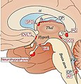
Español: Figura 1. A) Localización de órganos circunventriculares (CVO) y plexo coroideo (CP) en una vista esquemática de la superficie medial del cerebro.
Los CVO representados desempeñan funciones homeostáticas por sus funciones secretoras y sensoriales. La neurohipófisis, la parte distal (el lóbulo neural), recibe las neurohormonas hipotalámicas vasopresina y oxcitocina secretadas en el plexo vascular superficial en la parte proximal . El plexo vascular profundo de la eminencia mediana (ME) cumple una función sensorial que media señales metabólicas al núcleo arqueado (Arc) del hipotálamo. El organum vasculosum of the lamina terminalis (OVLT) participa en la osmorregulación controlando la liberación de vasopresina y en el control de la producción cíclica de las hormonas gonadotrópicas hipofisarias. El órgano subfornical (SFO) participa en la regulación de la liberación de vasopresina y la inducción de la sensación de sed provocada por la deshidratación. El órgano subcomisural (SCO) puede facilitar el flujo del líquido cefalorraquídeo en el acueducto cerebral entre el tercer y cuarto ventrículo. La glándula pineal (PG) secreta melatonina y estabiliza el cronometraje del fotoperíodo. El área postrema (AP) está involucrada en la regulación cardiovascular y respiratoria, así como en el control del centro del vómito en la médula.
Date
Source Circumventricular Organs and Parasite Neurotropism: Neglected Gates to the Brain? Front. Immunol. 2018. https://doi.org/10.3389/fimmu.2018.02877
Author Marina Bentivoglio; Krister Kristensson; Martin E. Rottenberg

Licensing

w:en:Creative Commons
attribution
This file is licensed under the Creative Commons Attribution 4.0 International license.
You are free:
  • to share – to copy, distribute and transmit the work
  • to remix – to adapt the work
Under the following conditions:
  • attribution – You must give appropriate credit, provide a link to the license, and indicate if changes were made. You may do so in any reasonable manner, but not in any way that suggests the licensor endorses you or your use.

Captions

Add a one-line explanation of what this file represents

Items portrayed in this file

depicts

11 December 2018

image/jpeg

8830e20a63d8a62da8fb5c153592d787cf548cd4

92,025 byte

480 pixel

461 pixel

File history

Click on a date/time to view the file as it appeared at that time.

Date/TimeThumbnailDimensionsUserComment
current15:02, 18 March 2022Thumbnail for version as of 15:02, 18 March 2022461 × 480 (90 KB)Sanador2.0Uploaded a work by Marina Bentivoglio; Krister Kristensson; Martin E. Rottenberg from Circumventricular Organs and Parasite Neurotropism: Neglected Gates to the Brain? Front. Immunol. 2018. https://doi.org/10.3389/fimmu.2018.02877 with UploadWizard

The following page uses this file:

Global file usage

The following other wikis use this file:

Metadata