File:Dracoraptor premaxillae.png

Original file (2,300 × 2,394 pixels, file size: 5.02 MB, MIME type: image/png)

Summary

Description

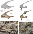
Details of Dracoraptor hanigani holotype skeleton National Museum Wales (NMW) 2015.5G.1–2015.5G.11.
Left: Right premaxilla in medial view. Top, simplified outline diagram highlighting the position of an elongate sub-narial vacuity. Middle, the premaxilla with matrix digitally removed. Bottom, premaxilla in situ.

Right: Left premaxilla with tooth in lateral view. Top, simplified outline diagram highlighting a series of? sensory openings at the tip of the element. Middle, the premaxilla and tooth with matrix digitally removed. Bottom, premaxilla in situ.
Date
Source http://journals.plos.org/plosone/article?id=10.1371/journal.pone.0145713
Author David M. Martill, Steven U. Vidovic, Cindy Howells, John R. Nudds

Licensing

w:en:Creative Commons
attribution
This file is licensed under the Creative Commons Attribution 2.5 Generic license.
You are free:
  • to share – to copy, distribute and transmit the work
  • to remix – to adapt the work
Under the following conditions:
  • attribution – You must give appropriate credit, provide a link to the license, and indicate if changes were made. You may do so in any reasonable manner, but not in any way that suggests the licensor endorses you or your use.
PLOS
PLOS
This file was published in a Public Library of Science journal. Their website states that the content of all PLOS journals is published under the Creative Commons Attribution 4.0 license (or its previous version depending on the publication date), unless indicated otherwise.

Captions

Add a one-line explanation of what this file represents

Items portrayed in this file

depicts

File history

Click on a date/time to view the file as it appeared at that time.

Date/TimeThumbnailDimensionsUserComment
current22:18, 22 January 2016Thumbnail for version as of 22:18, 22 January 20162,300 × 2,394 (5.02 MB)FunkMonk== {{int:filedesc}} == {{Information |Description=Left: Right premaxilla in medial view. Top, simplified outline diagram highlighting the position of an elongate sub-narial vacuity. Middle, the premaxilla with matrix digitally removed. Bottom, premaxil...

The following page uses this file:

Global file usage

The following other wikis use this file:

Metadata