Size of this preview: 535 × 599 pixels. Other resolutions: 214 × 240 pixels | 429 × 480 pixels | 686 × 768 pixels | 1,200 × 1,344 pixels.
Original file (1,200 × 1,344 pixels, file size: 212 KB, MIME type: image/jpeg)
File history
Click on a date/time to view the file as it appeared at that time.
| Date/Time | Thumbnail | Dimensions | User | Comment | |
|---|---|---|---|---|---|
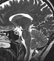
| current | 11:46, 10 September 2007 |  | 1,200 × 1,344 (212 KB) | Filip em | {{Information |Description=A T2-weighted sagittal MRI scan, from a patient with Chiari-like symptomatology, demonstrating tonsillar herniation less than 3 mm |Source=Raymond F Sekula Jr. et al. Dimensions of the posterior fossa in patients symptomatic for |
File usage
The following 3 pages use this file:
Global file usage
The following other wikis use this file:
- Usage on ca.wikipedia.org
- Usage on es.wikipedia.org
- Usage on fi.wikipedia.org
- Usage on he.wikipedia.org
- Usage on hr.wikipedia.org
- Usage on id.wikipedia.org
- Usage on it.wikipedia.org
- Usage on ja.wikipedia.org
- Usage on mk.wikipedia.org
- Usage on outreach.wikimedia.org
- Usage on pl.wikipedia.org
- Usage on sr.wikipedia.org
- Usage on www.wikidata.org