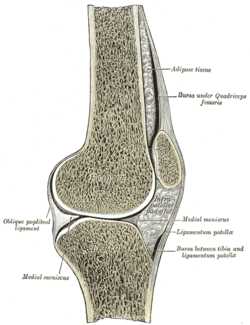
The knee bursae are the fluid-filled sacs and synovial pockets that surround and sometimes communicate with the knee joint cavity. The bursae are thin-walled, and filled with synovial fluid. They represent the weak point of the joint, but also provide enlargements to the joint space.[1] They can be grouped into either communicating and non-communicating bursae or, after their location – frontal, lateral, or medial.
| Knee bursae | |
|---|---|
 Sagittal section of right knee-joint, thus showing only frontal bursae. | |
| Anatomical terminology |
Frontal
editIn front, there are five bursae:
- the suprapatellar bursa or recess between the anterior surface of the lower part of the femur and the deep surface of the quadriceps femoris.[2] It allows for movement of the quadriceps tendon over the distal end of the femur. In about 85% of individuals, this bursa communicates with the knee joint. A distension of this bursa is therefore generally an indication of knee effusion.[3]
- the prepatellar bursa between the patella and the skin[2] It allows movement of the skin over the underlying patella.
- the deep infrapatellar bursa between the upper part of the tibia and the patellar ligament.[2] It allows for movement of the patellar ligament over the tibia.[4]
- the subcutaneous (or superficial) infrapatellar bursa between the patellar ligament and skin.[2]
- the pretibial bursa between the tibial tuberosity and the skin.[2] It allows for movement of the skin over the tibial tuberosity.[4]
Lateral
editLaterally there are four bursae:[2]
- the lateral gastrocnemius (subtendinous) bursa between the lateral head of the gastrocnemius and the joint capsule
- the fibular bursa between the lateral (fibular) collateral ligament and the tendon of the biceps femoris
- the fibulopopliteal bursa between the fibular collateral ligament and the tendon of the popliteus
- and the subpopliteal recess (or bursa) between the tendon of the popliteus and the lateral condyle of the femur
Medial
editMedially, there are five bursae:
- the medial gastrocnemius (subtendinous) bursa between the medial head of the gastrocnemius and the joint capsule[2]
- the anserine bursa between the medial (tibial) collateral ligament and the pes anserinus – the conjoined tendons of the sartorius, gracilis, and semitendinosus muscles.[2]
- the bursa semimembranosa between the medial collateral ligament and the tendon of the semimembranosus[2]
- there is one between the tendon of the semimembranosus and the head of the tibia[5]
- and occasionally there is a bursa between the tendons of the semimembranosus and semitendinosus[5]
See also
editNotes
editReferences
edit- Burgener, Francis A.; Meyers, Steven P.; Tan, Raymond K. (2002). Differential Diagnosis in Magnetic Resonance Imaging. Thieme. ISBN 1-58890-085-1.
- Cipriano, Joseph J. (2002). Photographic Manual of Regional Orthopaedic and Neurological Tests. Lippincott Williams & Wilkins. ISBN 0-7817-3552-1.
- "Gray's Anatomy (1918): The Knee-joint". Retrieved 26 April 2017.
- Platzer, Werner (2004). Color Atlas of Human Anatomy, Vol. 1: Locomotor System (5th ed.). Thieme. ISBN 3-13-533305-1.