Brodmann area 8 is one of Brodmann's cytologically defined regions of the brain. It is involved in planning complex movements.[citation needed]
| Brodmann area 8 | |
|---|---|
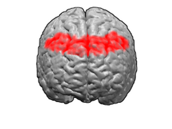
 Image of brain with Brodmann area 8 shown in red | |
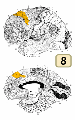
 Image of brain with Brodmann area 8 shown in orange | |
| Details | |
| Identifiers | |
| Latin | area frontalis intermedia |
| NeuroNames | 1034 |
| NeuroLex ID | birnlex_1739 |
| FMA | 68605 |
| Anatomical terms of neuroanatomy | |

Human
editBrodmann area 8, or BA8, is part of the frontal cortex in the human brain. Situated just anterior to the premotor cortex (BA6), it includes the frontal eye fields (so-named because they are believed to play an important role in the control of eye movements). Damage to this area, by stroke, trauma or infection, causes tonic deviation of the eyes towards the side of the injury. This finding occurs during the first few hours of an acute event such as cerebrovascular infarct (stroke) or hemorrhage (bleeding).
Guenon
editThe term Brodmann area 8 refers to a cytoarchitecturally defined portion of the frontal lobe of the guenon. Located rostral to the arcuate sulcus, it was not considered by Brodmann-1909 to be topographically homologous to the intermediate frontal area 8 of the human.
Distinctive features (Brodmann-1905): compared to Brodmann area 6-1909, area 8 has a diffuse but clearly present internal granular layer (IV); sublayer 3b of the external pyramidal layer (III) has densely distributed medium-sized pyramidal cells; the internal pyramidal layer (V) has larger ganglion cells densely distributed with some granule cells interspersed; the external granular layer (II) is denser and broader; cell layers are more distinct; the abundance of cells is somewhat greater.[1]
Functions
editThe area is involved in eye movements and possibly in the management of uncertainty. A functional magnetic resonance imaging study demonstrated that Brodmann area 8 activation occurs when test subjects experience uncertainty, and that with increasing uncertainty there is increasing activation.[2]
An alternative interpretation is that this activation in the frontal cortex encodes hope, a higher-order expectation positively correlated with uncertainty.[3]
Image
edit-
Animation.
-
front view.
-
Lateral view.
-
Medial view.
See also
editReferences
edit- ^ This article incorporates text available under the CC BY 3.0 license. "BrainInfo". Archived from the original on December 7, 2013. Retrieved December 3, 2013.
{{cite web}}: CS1 maint: bot: original URL status unknown (link) - ^ Volz KG, Schubotz RI, von Cramon DY (2005). "Variants of uncertainty in decision-making and their neural correlates". Brain Res. Bull. 67 (5): 403–12. doi:10.1016/j.brainresbull.2005.06.011. PMID 16216687. S2CID 15845324.
- ^ Chew, Soo Hong; Ho, Joanna L. (May 1994). "Hope: An empirical study of attitude toward the timing of uncertainty resolution". Journal of Risk and Uncertainty. 8 (3): 267–288. doi:10.1007/BF01064045. ISSN 0895-5646. JSTOR 41760728.