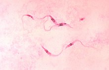
Kinetoplastida (or Kinetoplastea, as a class) is a group of flagellated protists belonging to the phylum Euglenozoa,[3][4] and characterised by the presence of a distinctive organelle called the kinetoplast (hence the name), a granule containing a large mass of DNA. The group includes a number of parasites responsible for serious diseases in humans and other animals, as well as various forms found in soil and aquatic environments. The organisms are commonly referred to as "kinetoplastids" or "kinetoplasts".[5]
| Kinetoplastida | |
|---|---|

| |
| Trypanosoma cruzi parasites | |
| Scientific classification | |
| Domain: | Eukaryota |
| Phylum: | Euglenozoa |
| Subphylum: | Glycomonada |
| Class: | Kinetoplastea Honigberg, 1963 emend. Cavalier-Smith, 1981[1][2] |
| Subdivisions | |
| Synonyms | |
| |
The kinetoplastids were first defined by Bronislaw M. Honigberg in 1963 as the members of the flagellated protozoans.[1] They are traditionally divided into the biflagellate Bodonidae and uniflagellate Trypanosomatidae; the former appears to be paraphyletic to the latter. One family of kinetoplastids, the trypanosomatids, is notable as it includes several genera which are exclusively parasitic. Bodo is a typical genus within kinetoplastida, which also includes various common free-living species which feed on bacteria. Others include Cryptobia and the parasitic Leishmania.
Taxonomy
editHistory
editHonigberg created the taxonomic names Kinetoplastida and Kinetoplastea in 1963.[1] Since then there is no consensus on the use of either of the two as a definite taxon. Kinetoplastea is more widely used as the class,[6][7][8][9][10] while Kinetoplastida is mostly used to designate the order,[4][11][12][13] but is also used as a class.[3][14] Lynn Margulis, who initially accepted Kinetoplastida as an order in 1974, later placed it as a class.[15] Use of Kinetoplastida as an order also creates confusion as there is already an older name Trypanosomatida Kent, 1880, under which the kinetoplastids are most often placed.[16]
Classification
editKinetoplastida is divided into two subclasses - Metakinetoplastina and Prokinetoplastina.[17][18]
- Family ?Bordnamonadidae Cavalier-Smith 2013
- Family ?Trypanophididae Poche 1911
- Subclass Prokinetoplastina Vickerman 2004
- Order Prokinetoplastida Vickerman 2004
- Family Ichthyobodonidae Isaksen et al. 2007
- Order Prokinetoplastida Vickerman 2004
- Subclass Metakinetoplastina Vickerman 2004
- Order Neobodonida Vickerman 2004
- Family Rhynchomonadidae Cavalier-Smith 2016
- Family Neobodonidae Cavalier-Smith 2016
- Order Parabodonida Vickerman 2004
- Family Parabodonidae Cavalier-Smith 2016 [Cryptobiaceae Poche 1911; Cryptobiidae Vickerman 1976; Trypanoplasmatidae Hartmann & Chagas 1910]
- Order Bodonida Hollande 1952 emend. Vickerman 1976
- Family Bodonidae Bütschli 1883
- Order Trypanosomatida Kent 1880 stat. n. Hollande 1952 emend. Vickerman 2004
- Family Trypanosomatidae Doflein 1901 [Trypanomorphidae Woodcock 1906]
- Order Neobodonida Vickerman 2004
Morphology
editKinetoplastids are eukaryotic and possess normal eukaryotic organelles, for example the nucleus, mitochondrion, golgi apparatus and flagellum. Along with these universal structures, kinetoplastids have several distinguishing morphological features such as the kinetoplast, sub-pellicular microtubule array and paraflagellar rod.[citation needed]
Mitochondrion and kinetoplast DNA
editThe kinetoplast, after which the class is named, is a dense DNA-containing granule within the cell's single mitochondrion, containing many copies of the mitochondrial genome. The structure is made up of a network of concatenated circular DNA molecules and their related structural proteins along with DNA and RNA polymerases. The kinetoplast is found at the base of a cell's flagella and is associated to the flagellum basal body by a cytoskeletal structure.[citation needed]
Cytoskeleton
editThe cytoskeleton of kinetoplastids is primarily made up of microtubules. These make a highly regular array, the sub-pellicular array, which runs parallel just under the cell surface along the long axis of the cell. Other microtubules with more specialised roles, such as the rootlet microtubules, are also present. Kinetoplastids are capable of forming actin microfilaments but their role in the cytoskeleton is not clear. Other cytoskeletal structures include the specialised attachment between the flagellum and the kinetoplast.[citation needed]
Flagella
editAll kinetoplastids possess at least one flagellum; species in the order trypanosomatida have one and bodonida have two. In kinetoplastids with two flagella most forms have a leading and trailing flagellum, the latter of which may be attached to the side of the cell. The flagella are used for locomotion and attachment to surfaces. The bases of the flagella are found in a specialised pocket structure which is also the location of the cytostome.[citation needed]
- Flagellum
- Flagellar membrane
- Flagellar axoneme
- Paraflagellar rod
- Flagellar attachment zone
- Flagellar-associated ER
- Pelicular microtubules
- Endosome, sorts material
- Glycosome
- Acidocalcisome
- Mitochondrion, creates ATP (energy) for the cell (discoid cristae)
- Nucleus
- Golgi apparatus, modifies proteins and sends them out of the cell
- Flagellar pocket collar
- Flagellar pocket
- Basal bodies
- Connecting fibres
- Kinetoplast, DNA-containing granule
- Antipodal site
Life cycle
editKinetoplastids may be free-living or parasitic. The order trypanosomatida is notable as it includes many genera which are exclusively parasitic. Trypanosomatids may have simple life cycles in a single host or more complex ones which progress through multiple differentiation stages in two hosts. Dramatic morphological changes are possible between lifecycle stages. Diseases caused by members of the order trypanosomatida include sleeping sickness and Chagas disease, caused by species of Trypanosoma, and leishmaniasis, caused by species of Leishmania.[19]
Trypanosoma brucei can undergo meiosis as a likely part of a sexual cycle.[20][21] Leishmania major is also capable of a meiotic process that is likely part of a sexual cycle.[22]
Gallery
edit-
Cryptobia sp.
-
Bodo sp.
-
Trypanosoma sp.
References
edit- ^ a b c Honigberg, B. M. (1963). "A contribution to systematics of the non-pigmented flagellates.". In Ludvík, J.; Lom, J.; Vávra J. (eds.). Progress in Protozoology: proceedings of the first International Congress on protozoology held at Prague. Academic Press.
- ^ Cavalier-Smith, T. (1981). Eukaryote kingdoms: seven or nine? Biosystems 14, 461–481.
- ^ a b Berman, Jules J. (2012). Taxonomic Guide to Infectious Diseases: Understanding the Biologic Classes of Pathogenic Organisms. London: Elsevier/Academic Press. p. 96. ISBN 978-0-12415-895-5.
- ^ a b "Kinetoplastida (kinetoplasts)". UniProt Consortium. Retrieved 22 January 2015.
- ^ Lukes, Julius (2009). "Kinetoplastida". The Tree of Life Web Project. Retrieved 10 September 2013.
- ^ "Kinetoplastea". EOL. Retrieved 22 January 2015.
- ^ "Kinetoplastea". World Register of Marine Species. Retrieved 22 January 2015.
- ^ "Kinetoplastea". ZipcodeZoo. Archived from the original on 22 January 2015. Retrieved 22 January 2015.
- ^ "Taxon: Class Kinetoplastea". The Taxonomicon. Archived from the original on 22 January 2015. Retrieved 22 January 2015.
- ^ Moreira, D. (2004). "An updated view of kinetoplastid phylogeny using environmental sequences and a closer outgroup: proposal for a new classification of the class Kinetoplastea". International Journal of Systematic and Evolutionary Microbiology. 54 (5): 1861–1875. doi:10.1099/ijs.0.63081-0. PMID 15388756.
- ^ "Kinetoplastida". EOL. Retrieved 22 January 2015.
- ^ "Kinetoplastida". Metalife. Archived from the original on 22 January 2015. Retrieved 22 January 2015.
- ^ "Kinetoplastida". NCBI Taxonomy. Retrieved 22 January 2015.
- ^ Gutierrez, Yezid (2000). Diagnostic Pathology of Parasitic Infections with Clinical Correlations (2 ed.). New York: Oxford University Press. p. 63. ISBN 978-0-1951214-38.
- ^ Chapman, Lynn Margulis, Michael J. (2009). Kingdoms & Domains : An Illustrated Guide to the Phyla of Life on Earth (4 ed.). Amsterdam: Academic Press/Elsevier. p. 158. ISBN 978-0-12-373621-5.
{{cite book}}: CS1 maint: multiple names: authors list (link) - ^ Deschamps, P.; Lara, E.; Marande, W.; Lopez-Garcia, P.; Ekelund, F.; Moreira, D. (2010). "Phylogenomic analysis of kinetoplastids supports that trypanosomatids arose from within bodonids". Molecular Biology and Evolution. 28 (1): 53–58. doi:10.1093/molbev/msq289. PMID 21030427.
- ^ Moreira, D; López-García, P; Vickerman, K (2004). "An updated view of kinetoplastid phylogeny using environmental sequences and a closer outgroup: proposal for a new classification of the class Kinetoplastea". International Journal of Systematic and Evolutionary Microbiology. 54 (Pt 5): 1861–1875. doi:10.1099/ijs.0.63081-0. PMID 15388756.
- ^ Stevens, JR (2008). "Kinetoplastid phylogenetics, with special reference to the evolution of parasitic trypanosomes". Parasite. 15 (3): 226–232. doi:10.1051/parasite/2008153226. PMID 18814685.
- ^ Scheckenbach F, Wylezich C, Mylnikov AP, Weitere M, Arndt H (October 2006). "Molecular comparisons of freshwater and marine isolates of the same morphospecies of heterotrophic flagellates". Appl Environ Microbiol. 72 (10): 6638–6643. Bibcode:2006ApEnM..72.6638S. doi:10.1128/AEM.02547-05. PMC 1610283. PMID 17021215.
{{cite journal}}: CS1 maint: date and year (link) - ^ Peacock L, Ferris V, Sharma R, Sunter J, Bailey M, Carrington M, Gibson W (2011). "Identification of the meiotic life cycle stage of Trypanosoma brucei in the tsetse fly" (PDF). Proc. Natl. Acad. Sci. U.S.A. 108 (9): 3671–6. Bibcode:2011PNAS..108.3671P. doi:10.1073/pnas.1019423108. PMC 3048101. PMID 21321215.
- ^ Gibson W (2015). "Liaisons dangereuses: sexual recombination among pathogenic trypanosomes". Res. Microbiol. 166 (6): 459–66. doi:10.1016/j.resmic.2015.05.005. hdl:1983/1ecb5cba-da25-4e93-a3cb-b00a0477cb23. PMID 26027775.
- ^ Akopyants NS, Kimblin N, Secundino N, Patrick R, Peters N, Lawyer P, Dobson DE, Beverley SM, Sacks DL (2009). "Demonstration of genetic exchange during cyclical development of Leishmania in the sand fly vector". Science. 324 (5924): 265–8. Bibcode:2009Sci...324..265A. doi:10.1126/science.1169464. PMC 2729066. PMID 19359589.
Bibliography
edit- Lumsden, W.H.R. & D.A. Evans (eds.). 1976-1979. Biology of the Kinetoplastida, 2 vols. London: Academic Press.