This article needs additional citations for verification. (April 2012) |
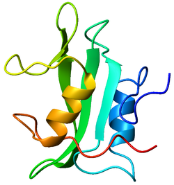
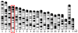
Tyrosin-protein kinase Lck (or lymphocyte-specific protein tyrosine kinase) is a 56 kDa protein that is found inside lymphocytes and encoded in the human by the LCK gene.[5] The Lck is a member of Src kinase family (SFK) and is important for the activation of T-cell receptor (TCR) signaling in both naive T cells and effector T cells. The role of Lck is less prominent in the activation or in the maintenance of memory CD8 T cells in comparison to CD4 T cells. In addition, the constitutive activity of the mouse Lck homolog varies among memory T cell subsets. It seems that in mice, in the effector memory T cell (TEM) population, more than 50% of Lck is present in a constitutively active conformation, whereas less than 20% of Lck is present as active form in central memory T cells. These differences are due to differential regulation by SH2 domain–containing phosphatase-1 (Shp-1) and C-terminal Src kinase.[6]
Lck is responsible for the initiation of the TCR signaling cascade inside the cell by phosphorylating immunoreceptor tyrosine-based activation motifs (ITAM) within the TCR-associated chains.
Lck can be found in different forms in immune cells: free in the cytosol or bound to the plasma membrane (PM) through myristoylation and palmitoylation. Due to the presence of the conserved CxxC motif (C20 and C23) in the zinc clasp structure, Lck is able to bind the cell surface coreceptors CD8 and\or CD4.
Bound and free Lck have different properties: free Lck has more pronounced kinase activity in comparison to bound Lck, and moreover, the free form produces a higher level of T cell activation.[7] The reasons for these differences are not well understood yet.
T cell signaling
editLck is most commonly found in T cells. It associates with the cytoplasmic tails of the CD4 and CD8 co-receptors on T helper cells and cytotoxic T cells,[8][9] respectively, to assist signaling from the T cell receptor (TCR) complex. T cells are able to respond to pathogen and cancer using T-cell receptor, nevertheless, they can also react to self-antigen causing the onset of autoimmune diseases. The T cells maturation occurs in the thymus and it is regulated by a threshold that defines the limit between the positive and the negative selection of thymocytes. in order to avoid the onset of autoimmune diseases, highly self-reactive T cells are removed during the negative selection, whereas, an amount of weak self-reactive T cells is required to promote an efficient immune response, therefore during the positive selection these cells are chosen for maturation. The threshold for positive and negative selection of developing T cells is regulated by the bound between the Lck and co-receptors.[10]
There are two main pools of T cells which mediate adaptive immune responses: CD4+ T cells (or helper T cells), and CD8+ T-cells (or cytotoxic T cells) which are MHCII-and MHCI restricted respectively. Despite their role in the immune system is different their activation is similar. Cytotoxic T cells are directly involved in the individuation and in the removal of infected cells, whereas helper T cells modulate other immune cells to supply the response.[11]
The initiation of immune response takes place when T cells encounter and recognize their cognate antigen. The antigen-presenting cells (APC) expose on their surface a fraction of the antigen that is recognized either from CD8+ T cells or CD4+ T cells. This binding leads to the activation of TCR signaling cascade in which the immunoreceptor tyrosine-based activation motifs (ITAM) located in the CD3-zeta chains (ζ-chains) of the TCR complex, are phosphorylated by Lck and less extended by Fyn.[12] Both coreceptor-bound and free Lck can phosphorylate the CD3 chains upon TCR activation, evidences suggest that the free form of Lck can be recruited and trigger the TCR signal faster than the coreceptor-bound Lck [7] Additionally, upon T cell activation, a fraction of kinase active Lck, translocate from outside of lipid rafts (LR) to inside lipid rafts where it interacts with and activates LR-resident Fyn, which is involved in further downstream signaling activation.[13][14] Once ITAM complex is phosphorylated the CD3 chains can be bound by another cytoplasmic tyrosine kinase called ZAP-70. In the case of CD8+ T cells, once ZAP70 binds CD3, the coreceptor associated with Lck binds the MHC stabilizing the TCR-MHC-peptide interaction. The phosphorylated form of ZAP-70 recruits another molecule in the signaling cascade called LAT (Linker for activation of T cells), a transmembrane protein. LAT acts as a scaffold able to regulate the TCR proximal signals in a phosphorylation-dependent manner.[15] The most important proteins recruited by phosphorylated LAT are Shc-Grb2-SOS, PI3K, and phospholipase C (PLC). The residue responsible for the recruitment of phospholipase C-γ1 (PLC-γ1) is Y132. This binding leads to the Tec family kinase ITK-mediated PLC-γ1 phosphorylation and activation that consequentially produce calcium (Ca2+) ions mobilization, and activation of important signaling cascades within the lymphocyte. These include the Ras-MEK-ERK pathway, which goes on to activate certain transcription factors such as NFAT, NF-κB, and AP-1. These transcription factors regulate the production of a plethora of gene products, most notable, cytokines such as Interleukin-2 that promote long-term proliferation and differentiation of the activated lymphocytes. In addition to the significance of Lck and Fyn in T cell receptor signaling, these two src kinases have also been shown to be important in TLR-mediated signaling in T cells.[16]
The function of Lck has been studied using several biochemical methods, including gene knockout (knock-out mice), Jurkat cells deficient in Lck (JCaM1.6), and siRNA-mediated RNA interference.
Lck activity regulation
editThe activity of the Lck can be positively or negatively regulated by the presence of other proteins such as the membrane protein CD146, the transmembrane tyrosine phosphatase CD45 and C-terminal Src kinase (Csk). In mice, CD146 directly interacts with the SH3 domain of coreceptor-free LCK via its cytoplasmic domain, promoting the LCK autophosphorylation.[17] There is very little understanding of the role of CD45 isoforms, it is known that they are cell type-specific, and that they depend on the state of activation and differentiation of cells. In naïve T cells in humans, CD45RA isoform is more frequent, whereas when cells are activated the CD45R0 isoform is expressed in higher concentrations. Mice express low levels of high molecular weight isoforms (CD45RABC) in thymocytes or peripheral T cells. Low levels of CD45RB are typical in primed cells, while high levels of CD45RB are found in both naïve and primed cells.[18] In general, CD45 acts to promote the active form of LCK by dephosphorylating a tyrosine (Y192) in its inhibitory C-terminal tail. The consequent trans-autophosphorylation of the tyrosine in the lck activation loop (Y394), stabilizes its active form promoting its open conformation[19] which further enhances the kinase activity and substrate binding. The Dephosphorylation of the Y394 site can also be regulated by SH2 domain-containing phosphatase 1 (SHP-1), PEST-domain enriched tyrosine phosphatase (PEP), and protein tyrosine phosphatase-PEST.[7] In contrast, Csk has an opposite role to that of CD45, it phosphorylated the Y505 of Lck promoting the closed conformation with inhibited kinase activity. When both Y394 and Y505 are unphosphorylated the lck show a basal kinase activity, vice versa, when phosphorylated, lck show similar activity to the Y394 single phosphorylated Lck [7]
Structure
editLck is a 56-kilodalton protein. The N-terminal tail of Lck is myristoylated and palmitoylated, which tethers the protein to the plasma membrane of the cell. The protein furthermore contains a SH3 domain, a SH2 domain and in the C-terminal part the tyrosine kinase domain. The two main phosphorylation sites on Lck are tyrosines 394 and 505. The former is an autophosphorylation site and is linked to activation of the protein. The latter is phosphorylated by Csk, which inhibits Lck because the protein folds up and binds its own SH2 domain. Lck thus serves as an instructive example that protein phosphorylation may result in both activation and inhibition.
Lck and disease
editMutations in Lck are liked to a various range of diseases such as SCID (Severe combined immunodeficiency) or CIDs. In these pathologies, the dysfunctional activation of the lck leads to T cell activation failure. Many pathologies are linked to the overexpression of Lck such as cancer, asthma, diabetes 1, rheumatoid arthritis, psoriasis, systemic lupus erythematosus, inflammatory bowel diseases (Crohn's disease and ulcerative colitis), organ graft rejection, atherosclerosis, hypersensitivity reactions, polyarthritis, dermatomyositis. The increase of the lck in colonic epithelial cells can lead to colorectal cancer. The lck play a role also in the Thymoma, an auto-immune disorder which involve thymus. Tumorigenesis is enhanced by abnormal proliferation of immature thymocytes due to low levels of Lck.[20]
Lymphoid protein tyrosine phosphatase (lyp), is one of the suppressor of lck activity and mutations in this protein are correlated with the onset of diabetes 1. Increased activity of lck promote the onset of the diabetes 1.
Regarding respiratory diseases, asthma is associated with the activation of th2 type of t cell whose differentiation is mediated by lck.[21] Moreover, mice with an unbalanced amount of lck show altered lung function which can consequentially leads to the onset of asthma. [22]
Substrates
editLck tyrosine phosphorylates a number of proteins, the most important of which are the CD3 receptor, CEACAM1, ZAP-70, SLP-76, the IL-2 receptor, Protein kinase C, ITK, PLC, SHC, RasGAP, Cbl, Vav1, and PI3K.
Inhibition
editIn resting T cells, Lck is constitutively inhibited by Csk phosphorylation on tyrosine 505. Lck is also inhibited by SHP-1 dephosphorylation on tyrosine 394. Lck can also be inhibited by Cbl ubiquitin ligase, which is part of the ubiquitin-mediated pathway.[23]
Saractinib, a specific inhibitor of LCK impairs maintenance of human T-ALL cells in vitro as well as in vivo by targeting this tyrosine kinase in cells displaying high level of lipid rafts.[24]
Masitinib also inhibits Lck, which may have some impact on its therapeutic effects in canine mastocytoma.[25]
HSP90 inhibitor NVP-BEP800 has been described to affect stability of the LCK kinase and growth of T-cell acute lymphoblastic leukemias.[26]
Interactions
editLck has been shown to interact with:
See also
editReferences
edit- ^ a b c GRCh38: Ensembl release 89: ENSG00000182866 – Ensembl, May 2017
- ^ a b c GRCm38: Ensembl release 89: ENSMUSG00000000409 – Ensembl, May 2017
- ^ "Human PubMed Reference:". National Center for Biotechnology Information, U.S. National Library of Medicine.
- ^ "Mouse PubMed Reference:". National Center for Biotechnology Information, U.S. National Library of Medicine.
- ^ "UniProt". www.uniprot.org. Retrieved 11 May 2024.
- ^ Moogk D, Zhong S, Yu Z, Liadi I, Rittase W, Fang V, et al. (July 2016). "Constitutive Lck Activity Drives Sensitivity Differences between CD8+ Memory T Cell Subsets". Journal of Immunology. 197 (2): 644–654. doi:10.4049/jimmunol.1600178. PMC 4935560. PMID 27271569.
- ^ a b c d Wei Q, Brzostek J, Sankaran S, Casas J, Hew LS, Yap J, et al. (July 2020). "Lck bound to coreceptor is less active than free Lck". Proceedings of the National Academy of Sciences of the United States of America. 117 (27): 15809–15817. Bibcode:2020PNAS..11715809W. doi:10.1073/pnas.1913334117. PMC 7355011. PMID 32571924.
- ^ Rudd CE, Trevillyan JM, Dasgupta JD, Wong LL, Schlossman SF (July 1988). "The CD4 receptor is complexed in detergent lysates to a protein-tyrosine kinase (pp58) from human T lymphocytes". Proceedings of the National Academy of Sciences of the United States of America. 85 (14): 5190–5194. Bibcode:1988PNAS...85.5190R. doi:10.1073/pnas.85.14.5190. PMC 281714. PMID 2455897.
- ^ Barber EK, Dasgupta JD, Schlossman SF, Trevillyan JM, Rudd CE (May 1989). "The CD4 and CD8 antigens are coupled to a protein-tyrosine kinase (p56lck) that phosphorylates the CD3 complex". Proceedings of the National Academy of Sciences of the United States of America. 86 (9): 3277–3281. Bibcode:1989PNAS...86.3277B. doi:10.1073/pnas.86.9.3277. PMC 287114. PMID 2470098.
- ^ Lovatt M, Filby A, Parravicini V, Werlen G, Palmer E, Zamoyska R (November 2006). "Lck regulates the threshold of activation in primary T cells, while both Lck and Fyn contribute to the magnitude of the extracellular signal-related kinase response". Molecular and Cellular Biology. 26 (22): 8655–8665. doi:10.1128/MCB.00168-06. PMC 1636771. PMID 16966372.
- ^ Horkova V, Drobek A, Mueller D, Gubser C, Niederlova V, Wyss L, et al. (February 2020). "Dynamics of the Coreceptor-LCK Interactions during T Cell Development Shape the Self-Reactivity of Peripheral CD4 and CD8 T Cells". Cell Reports. 30 (5): 1504–1514.e7. doi:10.1016/j.celrep.2020.01.008. PMC 7003063. PMID 32023465.
- ^ Janeway C (2012). "Chapter 7: Lymphocyte Receptor Signaling". janeway's immunobiology 8th edition. New York: Garland Science. p. 268.
- ^ Filipp D, Zhang J, Leung BL, Shaw A, Levin SD, Veillette A, Julius M (May 2003). "Regulation of Fyn through translocation of activated Lck into lipid rafts". The Journal of Experimental Medicine. 197 (9): 1221–1227. doi:10.1084/jem.20022112. PMC 2193969. PMID 12732664.
- ^ Filipp D, Moemeni B, Ferzoco A, Kathirkamathamby K, Zhang J, Ballek O, et al. (September 2008). "Lck-dependent Fyn activation requires C terminus-dependent targeting of kinase-active Lck to lipid rafts". The Journal of Biological Chemistry. 283 (39): 26409–26422. doi:10.1074/jbc.M710372200. PMC 3258908. PMID 18660530.
- ^ Lo WL, Shah NH, Rubin SA, Zhang W, Horkova V, Fallahee IR, et al. (November 2019). "Slow phosphorylation of a tyrosine residue in LAT optimizes T cell ligand discrimination". Nature Immunology. 20 (11): 1481–1493. doi:10.1038/s41590-019-0502-2. PMC 6858552. PMID 31611699.
- ^ Sharma N, Akhade AS, Qadri A (April 2016). "Src kinases central to T-cell receptor signaling regulate TLR-activated innate immune responses from human T cells". Innate Immunity. 22 (3): 238–244. doi:10.1177/1753425916632305. PMID 26888964.
- ^ Duan H, Jing L, Jiang X, Ma Y, Wang D, Xiang J, et al. (November 2021). "CD146 bound to LCK promotes T cell receptor signaling and antitumor immune responses in mice". The Journal of Clinical Investigation. 131 (21). doi:10.1172/JCI148568. PMC 8553567. PMID 34491908.
- ^ Tchilian EZ, Dawes R, Hyland L, Montoya M, Le Bon A, Borrow P, Hou S, Tough D, Beverley PC (2004). "Altered CD45 isoform expression affects lymphocyte function in CD45 Tg mice". International Immunology. 16 (9): 1323–1332. doi:10.1093/intimm/dxh135. PMID 15302847.
- ^ Courtney AH, Shvets AA, Lu W, Griffante G, Mollenauer M, Horkova V, et al. (October 2019). "CD45 functions as a signaling gatekeeper in T cells". Science Signaling. 12 (604): eaaw8151. doi:10.1126/scisignal.aaw8151. PMC 6948007. PMID 31641081.
- ^ Singh PK, Kashyap A, Silakari O (December 2018). "Exploration of the therapeutic aspects of Lck: A kinase target in inflammatory mediated pathological conditions". Biomedicine & Pharmacotherapy. 108: 1565–1571. doi:10.1016/j.biopha.2018.10.002. PMID 30372858. S2CID 53111664.
- ^ Kemp KL, Levin SD, Bryce PJ, Stein PL (April 2010). "Lck mediates Th2 differentiation through effects on T-bet and GATA-3". Journal of Immunology. 184 (8): 4178–4184. doi:10.4049/jimmunol.0901282. PMC 4889130. PMID 20237292.
- ^ Pernis AB, Rothman PB (May 2002). "JAK-STAT signaling in asthma". The Journal of Clinical Investigation. 109 (10): 1279–1283. doi:10.1172/JCI15786. PMC 150988. PMID 12021241.
- ^ Rao N, Miyake S, Reddi AL, Douillard P, Ghosh AK, Dodge IL, et al. (March 2002). "Negative regulation of Lck by Cbl ubiquitin ligase". Proceedings of the National Academy of Sciences of the United States of America. 99 (6): 3794–3799. Bibcode:2002PNAS...99.3794R. doi:10.1073/pnas.062055999. PMC 122603. PMID 11904433.
- ^ Buffière A, Accogli T, Saint-Paul L, Lucchi G, Uzan B, Ballerini P, et al. (September 2018). "Saracatinib impairs maintenance of human T-ALL by targeting the LCK tyrosine kinase in cells displaying high level of lipid rafts". Leukemia. 32 (9): 2062–2065. doi:10.1038/s41375-018-0081-5. PMID 29535432. S2CID 3833020.
- ^ Gil da Costa RM (July 2015). "C-kit as a prognostic and therapeutic marker in canine cutaneous mast cell tumours: From laboratory to clinic". Veterinary Journal. 205 (1): 5–10. doi:10.1016/j.tvjl.2015.05.002. hdl:10216/103345. PMID 26021891.
- ^ Mshaik R, Simonet J, Georgievski A, Jamal L, Bechoua S, Ballerini P, et al. (March 2021). "HSP90 inhibitor NVP-BEP800 affects stability of SRC kinases and growth of T-cell and B-cell acute lymphoblastic leukemias". Blood Cancer Journal. 11 (3): 61. doi:10.1038/s41408-021-00450-2. PMC 7973815. PMID 33737511.
- ^ Poghosyan Z, Robbins SM, Houslay MD, Webster A, Murphy G, Edwards DR (February 2002). "Phosphorylation-dependent interactions between ADAM15 cytoplasmic domain and Src family protein-tyrosine kinases". The Journal of Biological Chemistry. 277 (7): 4999–5007. doi:10.1074/jbc.M107430200. PMID 11741929.
- ^ Bell GM, Fargnoli J, Bolen JB, Kish L, Imboden JB (January 1996). "The SH3 domain of p56lck binds to proline-rich sequences in the cytoplasmic domain of CD2". The Journal of Experimental Medicine. 183 (1): 169–178. doi:10.1084/jem.183.1.169. PMC 2192399. PMID 8551220.
- ^ Taher TE, Smit L, Griffioen AW, Schilder-Tol EJ, Borst J, Pals ST (February 1996). "Signaling through CD44 is mediated by tyrosine kinases. Association with p56lck in T lymphocytes". The Journal of Biological Chemistry. 271 (5): 2863–2867. doi:10.1074/jbc.271.5.2863. PMID 8576267.
- ^ Ilangumaran S, Briol A, Hoessli DC (May 1998). "CD44 selectively associates with active Src family protein tyrosine kinases Lck and Fyn in glycosphingolipid-rich plasma membrane domains of human peripheral blood lymphocytes". Blood. 91 (10): 3901–3908. doi:10.1182/blood.V91.10.3901. PMID 9573028.
- ^ Hawash IY, Hu XE, Adal A, Cassady JM, Geahlen RL, Harrison ML (April 2002). "The oxygen-substituted palmitic acid analogue, 13-oxypalmitic acid, inhibits Lck localization to lipid rafts and T cell signaling". Biochimica et Biophysica Acta (BBA) - Molecular Cell Research. 1589 (2): 140–150. doi:10.1016/s0167-4889(02)00165-9. PMID 12007789.
- ^ Foti M, Phelouzat MA, Holm A, Rasmusson BJ, Carpentier JL (February 2002). "p56Lck anchors CD4 to distinct microdomains on microvilli". Proceedings of the National Academy of Sciences of the United States of America. 99 (4): 2008–2013. Bibcode:2002PNAS...99.2008F. doi:10.1073/pnas.042689099. PMC 122310. PMID 11854499.
- ^ Marcus SL, Winrow CJ, Capone JP, Rachubinski RA (November 1996). "A p56(lck) ligand serves as a coactivator of an orphan nuclear hormone receptor". The Journal of Biological Chemistry. 271 (44): 27197–27200. doi:10.1074/jbc.271.44.27197. PMID 8910285.
- ^ Hanada T, Lin L, Chandy KG, Oh SS, Chishti AH (October 1997). "Human homologue of the Drosophila discs large tumor suppressor binds to p56lck tyrosine kinase and Shaker type Kv1.3 potassium channel in T lymphocytes". The Journal of Biological Chemistry. 272 (43): 26899–26904. doi:10.1074/jbc.272.43.26899. PMID 9341123.
- ^ a b Sade H, Krishna S, Sarin A (January 2004). "The anti-apoptotic effect of Notch-1 requires p56lck-dependent, Akt/PKB-mediated signaling in T cells". The Journal of Biological Chemistry. 279 (4): 2937–2944. doi:10.1074/jbc.M309924200. PMID 14583609.
- ^ Prasad KV, Kapeller R, Janssen O, Repke H, Duke-Cohan JS, Cantley LC, Rudd CE (December 1993). "Phosphatidylinositol (PI) 3-kinase and PI 4-kinase binding to the CD4-p56lck complex: the p56lck SH3 domain binds to PI 3-kinase but not PI 4-kinase". Molecular and Cellular Biology. 13 (12): 7708–7717. doi:10.1128/mcb.13.12.7708. PMC 364842. PMID 8246987.
- ^ Yu CL, Jin YJ, Burakoff SJ (January 2000). "Cytosolic tyrosine dephosphorylation of STAT5. Potential role of SHP-2 in STAT5 regulation". The Journal of Biological Chemistry. 275 (1): 599–604. doi:10.1074/jbc.275.1.599. PMID 10617656.
- ^ Chiang GG, Sefton BM (June 2001). "Specific dephosphorylation of the Lck tyrosine protein kinase at Tyr-394 by the SHP-1 protein-tyrosine phosphatase". The Journal of Biological Chemistry. 276 (25): 23173–23178. doi:10.1074/jbc.M101219200. PMID 11294838.
- ^ Lorenz U, Ravichandran KS, Pei D, Walsh CT, Burakoff SJ, Neel BG (March 1994). "Lck-dependent tyrosyl phosphorylation of the phosphotyrosine phosphatase SH-PTP1 in murine T cells". Molecular and Cellular Biology. 14 (3): 1824–1834. doi:10.1128/mcb.14.3.1824. PMC 358540. PMID 8114715.
- ^ Koretzky GA, Kohmetscher M, Ross S (April 1993). "CD45-associated kinase activity requires lck but not T cell receptor expression in the Jurkat T cell line". The Journal of Biological Chemistry. 268 (12): 8958–8964. doi:10.1016/S0021-9258(18)52965-3. PMID 8473339.
- ^ Ng DH, Watts JD, Aebersold R, Johnson P (January 1996). "Demonstration of a direct interaction between p56lck and the cytoplasmic domain of CD45 in vitro". The Journal of Biological Chemistry. 271 (3): 1295–1300. doi:10.1074/jbc.271.3.1295. PMID 8576115.
- ^ Gorska MM, Stafford SJ, Cen O, Sur S, Alam R (February 2004). "Unc119, a novel activator of Lck/Fyn, is essential for T cell activation". The Journal of Experimental Medicine. 199 (3): 369–379. doi:10.1084/jem.20030589. PMC 2211793. PMID 14757743.
- ^ a b Thome M, Duplay P, Guttinger M, Acuto O (June 1995). "Syk and ZAP-70 mediate recruitment of p56lck/CD4 to the activated T cell receptor/CD3/zeta complex". The Journal of Experimental Medicine. 181 (6): 1997–2006. doi:10.1084/jem.181.6.1997. PMC 2192070. PMID 7539035.
- ^ Oda H, Kumar S, Howley PM (August 1999). "Regulation of the Src family tyrosine kinase Blk through E6AP-mediated ubiquitination". Proceedings of the National Academy of Sciences of the United States of America. 96 (17): 9557–9562. Bibcode:1999PNAS...96.9557O. doi:10.1073/pnas.96.17.9557. PMC 22247. PMID 10449731.
- ^ Pelosi M, Di Bartolo V, Mounier V, Mège D, Pascussi JM, Dufour E, et al. (May 1999). "Tyrosine 319 in the interdomain B of ZAP-70 is a binding site for the Src homology 2 domain of Lck". The Journal of Biological Chemistry. 274 (20): 14229–14237. doi:10.1074/jbc.274.20.14229. PMID 10318843.
Further reading
edit- Sasaoka T, Kobayashi M (August 2000). "The functional significance of Shc in insulin signaling as a substrate of the insulin receptor". Endocrine Journal. 47 (4): 373–381. doi:10.1507/endocrj.47.373. PMID 11075717.
- Goldmann WH (2003). "p56(lck) Controls phosphorylation of filamin (ABP-280) and regulates focal adhesion kinase (pp125(FAK))". Cell Biology International. 26 (6): 567–571. doi:10.1006/cbir.2002.0900. PMID 12171035. S2CID 86450727.
- Mustelin T, Taskén K (April 2003). "Positive and negative regulation of T-cell activation through kinases and phosphatases". The Biochemical Journal. 371 (Pt 1): 15–27. doi:10.1042/BJ20021637. PMC 1223257. PMID 12485116.
- Zamoyska R, Basson A, Filby A, Legname G, Lovatt M, Seddon B (February 2003). "The influence of the src-family kinases, Lck and Fyn, on T cell differentiation, survival and activation". Immunological Reviews. 191: 107–118. doi:10.1034/j.1600-065X.2003.00015.x. PMID 12614355. S2CID 10156186.
- Summy JM, Gallick GE (December 2003). "Src family kinases in tumor progression and metastasis". Cancer and Metastasis Reviews. 22 (4): 337–358. doi:10.1023/A:1023772912750. PMID 12884910. S2CID 12380282.
- Leavitt SA, SchOn A, Klein JC, Manjappara U, Chaiken IM, Freire E (February 2004). "Interactions of HIV-1 proteins gp120 and Nef with cellular partners define a novel allosteric paradigm". Current Protein & Peptide Science. 5 (1): 1–8. doi:10.2174/1389203043486955. PMID 14965316.
- Tolstrup M, Ostergaard L, Laursen AL, Pedersen SF, Duch M (April 2004). "HIV/SIV escape from immune surveillance: focus on Nef". Current HIV Research. 2 (2): 141–151. doi:10.2174/1570162043484924. PMID 15078178.
- Palacios EH, Weiss A (October 2004). "Function of the Src-family kinases, Lck and Fyn, in T-cell development and activation". Oncogene. 23 (48): 7990–8000. doi:10.1038/sj.onc.1208074. PMID 15489916. S2CID 20109652.
- Joseph AM, Kumar M, Mitra D (January 2005). "Nef: "necessary and enforcing factor" in HIV infection". Current HIV Research. 3 (1): 87–94. doi:10.2174/1570162052773013. PMID 15638726.
- Levinson AD, Oppermann H, Levintow L, Varmus HE, Bishop JM (October 1978). "Evidence that the transforming gene of avian sarcoma virus encodes a protein kinase associated with a phosphoprotein". Cell. 15 (2): 561–572. doi:10.1016/0092-8674(78)90024-7. PMID 214242. S2CID 40461709.
- Thomas PM, Samelson LE (June 1992). "The glycophosphatidylinositol-anchored Thy-1 molecule interacts with the p60fyn protein tyrosine kinase in T cells". The Journal of Biological Chemistry. 267 (17): 12317–12322. doi:10.1016/S0021-9258(19)49841-4. PMID 1351058.
- Shenoy-Scaria AM, Kwong J, Fujita T, Olszowy MW, Shaw AS, Lublin DM (December 1992). "Signal transduction through decay-accelerating factor. Interaction of glycosyl-phosphatidylinositol anchor and protein tyrosine kinases p56lck and p59fyn 1". Journal of Immunology. 149 (11): 3535–3541. doi:10.4049/jimmunol.149.11.3535. PMID 1385527. S2CID 23189716.
- Weber JR, Bell GM, Han MY, Pawson T, Imboden JB (August 1992). "Association of the tyrosine kinase LCK with phospholipase C-gamma 1 after stimulation of the T cell antigen receptor". The Journal of Experimental Medicine. 176 (2): 373–379. doi:10.1084/jem.176.2.373. PMC 2119313. PMID 1500851.
- Cefai D, Ferrer M, Serpente N, Idziorek T, Dautry-Varsat A, Debre P, Bismuth G (July 1992). "Internalization of HIV glycoprotein gp120 is associated with down-modulation of membrane CD4 and p56lck together with impairment of T cell activation". Journal of Immunology. 149 (1): 285–294. doi:10.4049/jimmunol.149.1.285. PMID 1535086. S2CID 25896387.
- Soula M, Fagard R, Fischer S (February 1992). "Interaction of human immunodeficiency virus glycoprotein 160 with CD4 in Jurkat cells increases p56lck autophosphorylation and kinase activity". International Immunology. 4 (2): 295–299. doi:10.1093/intimm/4.2.295. PMID 1535787.
- Crise B, Rose JK (April 1992). "Human immunodeficiency virus type 1 glycoprotein precursor retains a CD4-p56lck complex in the endoplasmic reticulum". Journal of Virology. 66 (4): 2296–2301. doi:10.1128/JVI.66.4.2296-2301.1992. PMC 289024. PMID 1548763.
- Molina TJ, Kishihara K, Siderovski DP, van Ewijk W, Narendran A, Timms E, et al. (May 1992). "Profound block in thymocyte development in mice lacking p56lck". Nature. 357 (6374): 161–164. Bibcode:1992Natur.357..161M. doi:10.1038/357161a0. PMID 1579166. S2CID 4363506.
- Yoshida H, Koga Y, Moroi Y, Kimura G, Nomoto K (February 1992). "The effect of p56lck, a lymphocyte specific protein tyrosine kinase, on the syncytium formation induced by human immunodeficiency virus envelope glycoprotein". International Immunology. 4 (2): 233–242. doi:10.1093/intimm/4.2.233. PMID 1622897.
- Torigoe T, O'Connor R, Santoli D, Reed JC (August 1992). "Interleukin-3 regulates the activity of the LYN protein-tyrosine kinase in myeloid-committed leukemic cell lines". Blood. 80 (3): 617–624. doi:10.1182/blood.V80.3.617.617. PMID 1638019.
External links
edit- lck+Kinase at the U.S. National Library of Medicine Medical Subject Headings (MeSH)
- Overview of all the structural information available in the PDB for UniProt: P06239 (Tyrosine-protein kinase Lck) at the PDBe-KB.


