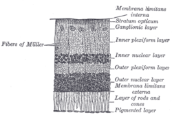
The retinal nerve fiber layer (RNFL) or nerve fiber layer, stratum opticum, is part of the anatomy of the eye.
| Retinal nerve fiber layer | |
|---|---|
 Section of retina. (Stratum opticum labeled at right, second from the top.) | |
 Plan of retinal neurons. (Stratum opticum labeled at left, second from the top.) | |
| Details | |
| Identifiers | |
| Latin | stratum neurofibrarum retinae |
| TA98 | A15.2.04.017 |
| FMA | 58688 |
| Anatomical terminology | |
Physical structure
editThe RNFL formed by the expansion of the fibers of the optic nerve; it is thickest near the optic disc, gradually diminishing toward the ora serrata.
As the nerve fibers pass through the lamina cribrosa sclerae they lose their medullary sheaths and are continued onward through the choroid and retina as simple axis-cylinders.
When they reach the internal surface of the retina they radiate from their point of entrance over this surface grouped in bundles, and in many places arranged in plexuses.
Most of the fibers are centripetal, and are the direct continuations of the axis-cylinder processes of the cells of the ganglionic layer, but a few of them are centrifugal and ramify in the inner plexiform and inner nuclear layers, where they end in enlarged extremities.
Measurement
editRNFL measurement can be made by Optical coherence tomography.[1]
Relation with diseases
editRNFL reduction
editRetinitis pigmentosa
editPatients with retinitis pigmentosa have abnormal thinning of the RNFL which correlates with the severity of the disease.[2] However the thickness of the RNFL also decreases with age and not visual acuity.[3] The sparing of this layer is important in the treatment of the disease as it is the basis for connecting retinal prostheses to the optic nerve, or implanting stem cells that could regenerate the lost photoreceptors.
Asymmetric RNFL
editRNFL asymmetry is the difference between the RNFL of the left and right eyes. In healthy patients, one study (2008, n=109) found asymmetry to be typically between 0-8μm, but occasionally higher, with average asymmetry of c.3μm at age 25 rising to 5μm at age 60.[4] A 2011 study (n=284) concluded that RNFL asymmetry exceeding 9μm may be considered statistically significant and may be indicative of early glaucomatous damage.[5] A 2023 study of 4034 children found mean RNFL of 106μm with SD of 9.4μm.[6]
Optic neuritis
editRNFL asymmetry has been proposed as a strong indicator of optic neuritis,[7][8] with one small study proposing that asymmetry of 5–6μm was "a robust structural threshold for identifying the presence of a unilateral optic nerve lesion in MS."[9] Optic neuritis is often associated with multiple sclerosis, and RNFL data may indicate the pace of future development of the MS.[10][11]
Glaucoma
editRNFL asymmetry may be produced by glaucoma.[12][13][14][15] Glaucoma is a lead cause of irreversible blindness. Resesrch in RNFL and optic nerve head (ONH) abnormalities may enable early detection and diagnosis of glaucoma.[2]
Correlation with ethnicity
editOther factors affecting RNFL
editSome processes can excite RNFL apoptosis. Harmful situations which can damage RNFL include high intraocular pressure, high fluctuation on phase of intraocular pressure, inflammation, vascular disease and any kind of hypoxia. Gede Pardianto (2009) reported 6 cases of RNFL thickness change after the procedures of phacoemulsification.[18] Sudden intraocular fluctuation in any kind of intraocular surgeries maybe harmful to RNFL in accordance with mechanical stress on sudden compression and also ischemic effect of micro emboly as the result of the sudden decompression that may generate micro bubble that can clog micro vessels.[19]
See also
editReferences
edit- ^ https://eyewiki.org/Optic_Nerve_and_Retinal_Nerve_Fiber_Imaging
- ^ a b Desissaire S, Pollreisz A, Sedova A, Hajdu D, Datlinger F, Steiner S, et al. (October 2020). "Analysis of retinal nerve fiber layer birefringence in patients with glaucoma and diabetic retinopathy by polarization sensitive OCT". Biomedical Optics Express. 11 (10): 5488–5505. doi:10.1364/BOE.402475. PMC 7587266. PMID 33149966.
- ^ Oishi A, Otani A, Sasahara M, Kurimoto M, Nakamura H, Kojima H, et al. (March 2009). "Retinal nerve fiber layer thickness in patients with retinitis pigmentosa". Eye. 23 (3): 561–6. doi:10.1038/eye.2008.63. PMID 18344951.
- ^ Budenz DL (2008). "Symmetry Between the Right and Left Eyes of the Normal Retinal Nerve Fiber Layer Measured with Optical Coherence Tomography (An AOS Thesis)". Transactions of the American Ophthalmological Society. 106: 252–275. PMC 2646446. PMID 19277241.
- ^ MWANZA JC, DURBIN MK, BUDENZ DL (2011). "Interocular Symmetry in Peripapillary Retinal Nerve Fiber Layer Thickness Measured With the Cirrus HD-OCT in Healthy Eyes". American Journal of Ophthalmology. 151 (3): 514–21.e1. doi:10.1016/j.ajo.2010.09.015. PMC 5457794. PMID 21236402.
- ^ Zhang XJ, Wang YM, Jue Z, Chan HN, Lau YH, Zhang W, et al. (2023). "Interocular Symmetry in Retinal Nerve Fiber Layer Thickness in Children: The Hong Kong Children Eye Study". Ophthalmology and Therapy. 12 (6): 3373–3382. doi:10.1007/s40123-023-00825-7. PMC 10640485. PMID 37851163.
- ^ Jiang H, Delgado S, Wang J (2021). "Advances in ophthalmic structural and functional measures in multiple sclerosis: do the potential ocular biomarkers meet the unmet needs?". Current Opinion in Neurology. 34 (1): 97–107. doi:10.1097/WCO.0000000000000897. PMC 7856092. PMID 33278142.
- ^ Nij Bijvank J, Uitdehaag BM, Petzold A (2021). "Short report: Retinal inter-eye difference and atrophy progression in multiple sclerosis diagnostics". Journal of Neurology, Neurosurgery, and Psychiatry. 93 (2): 216–219. doi:10.1136/jnnp-2021-327468. PMC 8785044. PMID 34764152.
- ^ Nolan RC, Galetta SL, Frohman TC, Frohman EM, Calabresi PA, Castrillo-Viguera C, et al. (December 2018). "Optimal Intereye Difference Thresholds in Retinal Nerve Fiber Layer Thickness for Predicting a Unilateral Optic Nerve Lesion in Multiple Sclerosis". Journal of Neuro-Ophthalmology. 38 (4): 451–458. doi:10.1097/WNO.0000000000000629. PMC 8845082. PMID 29384802.
- ^ Bsteh G, Hegen H, Altmann P, Auer M, Berek K, Pauli FD, et al. (October 19, 2020). "Retinal layer thinning is reflecting disability progression independent of relapse activity in multiple sclerosis". Multiple Sclerosis Journal: Experimental, Translational and Clinical. 6 (4): 2055217320966344. doi:10.1177/2055217320966344. PMC 7604994. PMID 33194221.
- ^ Martinez-Lapiscina EH, Arnow S, Wilson JA, Saidha S, Preiningerova JL, Oberwahrenbrock T, et al. (May 2016). "Retinal thickness measured with optical coherence tomography and risk of disability worsening in multiple sclerosis: a cohort study". The Lancet. Neurology. 15 (6): 574–584. doi:10.1016/S1474-4422(16)00068-5. PMID 27011339.
- ^ Rodríguez-Robles F, Verdú-Monedero R, Berenguer-Vidal R, Morales-Sánchez J, Sellés-Navarro I (January 21, 2023). "Analysis of the Asymmetry between Both Eyes in Early Diagnosis of Glaucoma Combining Features Extracted from Retinal Images and OCTs into Classification Models". Sensors. 23 (10): 4737. Bibcode:2023Senso..23.4737R. doi:10.3390/s23104737. PMC 10220946. PMID 37430650.
- ^ Choplin NT, Craven ER, Reus NJ, Lemij HG, Barnebey H (January 2015). "21 - Retinal Nerve Fiber Layer (RNFL) Photography and Computer Analysis". Retinal Nerve Fiber Layer (RNFL) Photography and Computer Analysis - ScienceDirect. W.B. Saunders. pp. 244–260. doi:10.1016/B978-0-7020-5193-7.00021-2. ISBN 978-0-7020-5193-7.
- ^ Berenguer-Vidal R, Verdú-Monedero R, Morales-Sánchez J, Sellés-Navarro I, Kovalyk O (May 31, 2022). Analysis of the Asymmetry in RNFL Thickness Using Spectralis OCT Measurements in Healthy and Glaucoma Patients. Lecture Notes in Computer Science. Vol. 13258. Springer-Verlag. pp. 507–515. doi:10.1007/978-3-031-06242-1_50. ISBN 978-3-031-06241-4 – via ACM Digital Library.
- ^ "RNFL Analysis in the Diagnosis of Glaucoma". Glaucoma Today.
- ^ Nousome D, McKean-Cowdin R, Richter GM, Burkemper B, Torres M, Varma R, et al. (2020). "Retinal Nerve Fiber Layer Thickness in Healthy Eyes of African, Chinese, and Latino Americans: A Population-based Multiethnic Study". Ophthalmology. 128 (7): 1005–1015. doi:10.1016/j.ophtha.2020.11.015. PMC 8128930. PMID 33217471.
- ^ Heidary F, Gharebaghi R, Wan Hitam WH, Shatriah I (September 2010). "Nerve fiber layer thickness". Ophthalmology. 117 (9): 1861–1862. doi:10.1016/j.ophtha.2010.05.024. PMID 20816254.
- ^ Pardianto G (2009). "Mastering phacoemulsification in Mimbar Ilmiah". Oftalmologi Indonesia. 10: 26.
- ^ Pardianto G, Moeloek N, Reveny J, Wage S, Satari I, Sembiring R, et al. (2013). "Retinal thickness changes after phacoemulsification". Clinical Ophthalmology. 7. Auckland, N.Z.: 2207–14. doi:10.2147/OPTH.S53223. PMC 3821754. PMID 24235812.
This article incorporates text in the public domain from page 1015 of the 20th edition of Gray's Anatomy (1918)
External links
edit- Histology image: 07902loa – Histology Learning System at Boston University