This article needs more reliable medical references for verification or relies too heavily on primary sources. (November 2021) |  |
Primary gastric lymphoma (lymphoma that originates in the stomach itself)[1] is an uncommon condition, accounting for less than 15% of gastric malignancies and about 2% of all lymphomas. However, the stomach is a very common extranodal site for lymphomas (lymphomas originate elsewhere and metastasise to the stomach).[2] It is also the most common source of lymphomas in the gastrointestinal tract.[3]
| Gastric lymphoma | |
|---|---|
 | |
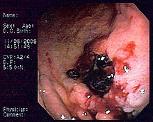
| Endoscopic image of gastric MALT lymphoma taken in body of stomach in patient who presented with upper GI hemorrhage. Appearance is similar to gastric ulcer with adherent blood clot. | |
| Specialty | Oncology |
Signs and symptoms
editSymptoms include epigastric pain, early satiety, fatigue and weight loss. Most people affected by primary gastric lymphoma are over 60 years old.[4]
Risk factors
editRisk factors for gastric lymphoma include the following:
- Helicobacter pylori[5]
- Long-term immunosuppressant drug therapy
- HIV infection
Pathophysiology
editThe majority of gastric lymphomas are non-Hodgkin's lymphoma of B-cell origin. These tumors may range from well-differentiated, superficial involvements (MALT) to high-grade, large-cell lymphomas. Sometimes, it's hard to differentiate poorly differentiated high grade B-cell gastric lymphoma from gastric adenocarcinoma clinically or radiologically, yet histopathology with immunohistochemistry is recommended to stain specific markers on the malignant cell that favor the diagnosis of lymphoma. Immunohistochemistry stains specific clusters of differentiation that are present on B-cells like CD20. Cytokeratin is also a surface marker that is presented on epithelial cells, is stained histochemically and favors the diagnosis of epithelial tumors like adenocarcinoma.
Distinguishing poorly differentiated gastric lymphoma from adenocarcinoma is essential because the prognosis and modalities of treatment differ significantly.
Other lymphomas involving the stomach include mantle cell lymphoma and T-cell lymphomas which may be associated with enteropathy; the latter usually occur in the small bowel but have been reported in the stomach.
Diagnosis
editThese lymphomas are difficult to differentiate from gastric adenocarcinoma. The lesions are usually ulcers with a ragged, thickened mucosal pattern on contrast radiographs.
The diagnosis is typically made by biopsy at the time of endoscopy. Several endoscopic findings have been reported, including solitary ulcers, thickened gastric folds, mass lesions and nodules. As there may be infiltration of the submucosa, larger biopsy forceps, endoscopic ultrasound guided biopsy, endoscopic submucosal resection, or laparotomy may be required to obtain tissue.
Imaging investigations including CT scans or endoscopic ultrasound are useful to stage disease. Hematological parameters are usually checked to assist with staging and to exclude concomitant leukemia. An elevated LDH level may be suggestive of lymphoma.
Treatment
editDiffuse large B-cell lymphomas of the stomach are primarily treated with chemotherapy with CHOP (cyclophosphamide+doxorubicine+vincristine+prednisone) with or without rituximab being a usual first choice.[citation needed]
Antibiotic treatment to eradicate H. pylori is indicated as first line therapy for MALT lymphomas. About 80% of MALT lymphomas completely regress with eradication therapy.[6] Radiation treatment for H. pylori–negative gastric malt lymphoma, has a high success rate, 90% or better after 5 years. Second line therapy for MALT lymphomas is usually chemotherapy with a single agent, and complete response rates of greater than 70% have been reported.[7]
Subtotal gastrectomy, with post-operative chemotherapy is undertaken in refractory cases, or in the setting of complications, including gastric outlet obstruction.
See also
editReferences
edit- ^ Dawson IM, Cornes JS, Morson BC (July 1961). "Primary malignant lymphoid tumours of the intestinal tract. Report of 37 cases with a study of factors influencing prognosis". Br J Surg. 49 (213): 80–9. doi:10.1002/bjs.18004921319. PMID 13884035. S2CID 43422925.
- ^ Aisenberg AC (October 1995). "Coherent view of non-Hodgkin's lymphoma". J. Clin. Oncol. 13 (10): 2656–75. doi:10.1200/JCO.1995.13.10.2656. PMID 7595720.[permanent dead link]
- ^ Koch P, del Valle F, Berdel WE, et al. (September 2001). "Primary gastrointestinal non-Hodgkin's lymphoma: I. Anatomic and histologic distribution, clinical features, and survival data of 371 patients registered in the German Multicenter Study GIT NHL 01/92". J. Clin. Oncol. 19 (18): 3861–73. doi:10.1200/JCO.2001.19.18.3861. PMID 11559724. Archived from the original on August 5, 2012.
- ^ Thirlby RC (January 1993). "Gastrointestinal lymphoma: a surgical perspective". Oncology (Williston Park, N.Y.). 7 (1): 29–32, 34, discussion 34, 37–8. PMID 8420540.
- ^ Parsonnet J, Hansen S, Rodriguez L, et al. (May 1994). "Helicobacter pylori infection and gastric lymphoma". N. Engl. J. Med. 330 (18): 1267–71. doi:10.1056/NEJM199405053301803. PMID 8145781.
- ^ Stathis, A; Bertoni, F; Zucca, E (September 2010). "Treatment of gastric marginal zone lymphoma of MALT type". Expert Opinion on Pharmacotherapy. 11 (13): 2141–52. doi:10.1517/14656566.2010.497141. PMID 20586708. S2CID 6796557.
- ^ Hammel P, Haioun C, Chaumette MT, et al. (October 1995). "Efficacy of single-agent chemotherapy in low-grade B-cell mucosa-associated lymphoid tissue lymphoma with prominent gastric expression". J. Clin. Oncol. 13 (10): 2524–9. doi:10.1200/JCO.1995.13.10.2524. PMID 7595703.[permanent dead link]