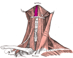
The geniohyoid muscle is a narrow paired muscle situated superior to the medial border of the mylohyoid muscle. It is named for its passage from the chin ("genio-" is a standard prefix for "chin")[1] to the hyoid bone.
| Geniohyoid muscle | |
|---|---|
 Anterior view. Geniohyoid muscle labeled at upper center left | |
 Extrinsic muscles of the tongue. Left side. | |
| Details | |
| Origin | Inferior mental spine of mandible |
| Insertion | Hyoid bone |
| Artery | Branches of the lingual artery. |
| Nerve | C1 via the hypoglossal nerve |
| Actions | Carry hyoid bone and the tongue upward during deglutition |
| Identifiers | |
| Latin | musculus geniohyoideus |
| TA98 | A04.2.03.007 |
| TA2 | 2166 |
| FMA | 46325 |
| Anatomical terms of muscle | |
Structure
editThe geniohyoid is a paired short muscle that arises from the inferior mental spine, on the back of the mandibular symphysis, and runs backward and slightly downward, to be inserted into the anterior surface of the body of the hyoid bone.[2]: 346 It lies in contact with its fellow of the opposite side. It thus belongs to the suprahyoid muscles. The muscle receives its blood supply from branches of the lingual artery.[3]
Innervation
editThe geniohyoid muscle is innervated by fibres from the first cervical spinal nerve travelling alongside the hypoglossal nerve.[2][4][5] Although the first three cervical nerves give rise to the ansa cervicalis, the geniohyoid muscle is said to be innervated by the first cervical nerve, as some of its efferent fibers do not contribute to ansa cervicalis.[clarification needed]
Variations
editIt may be blended with the one on opposite side or double; slips to greater cornu of hyoid bone and genioglossus occur.[citation needed]
Function
editThe geniohyoid muscle brings the hyoid bone forward and upwards.[2] This dilates the upper airway, assisting respiration.[4] During the first act of deglutition, when the mass of food is being driven from the mouth into the pharynx, the hyoid bone, and with it the tongue, is carried upward and forward by the anterior bellies of the Digastrici, the Mylohyoidei, and Geniohyoidei. It also assists in depressing the mandible.[3]
History
editThe inclined position of the geniohyoid muscle has been contrasted to the horizontal position in neanderthals.[6]
Additional images
edit-
Illustration of the hyoid bone showing the insertion point of the geniohyoid muscle
-
Sagittal section of nose mouth, pharynx, and larynx.
-
Geniohyoid muscle
-
Geniohyoid muscle
-
Geniohyoid muscle
See also
editReferences
editThis article incorporates text in the public domain from page 393 of the 20th edition of Gray's Anatomy (1918)
- ^ "Genio-". Merriam-Webster Dictionary.
Chin
- ^ a b c Singh I (2009). Essentials of anatomy (2nd ed.). New Delhi: Jaypee Bros. p. 346. ISBN 978-81-8448-461-8.[permanent dead link]
- ^ a b Drake RL, Vogel W, Mitchell AW (2020). "Head and Neck". Gray's Anatomy for Students (Fourth ed.). Philadelphia, PA. pp. 823–1121. ISBN 978-0-323-39304-1.
{{cite book}}: CS1 maint: location missing publisher (link) - ^ a b Takahashi S, Ono T, Ishiwata Y, Kuroda T (December 2002). "Breathing modes, body positions, and suprahyoid muscle activity". Journal of Orthodontics. 29 (4): 307–13, discussion 279. CiteSeerX 10.1.1.514.2998. doi:10.1093/ortho/29.4.307. PMID 12444272.
- ^ Drake RL, Vogl W, Mitchell AW, Gray H (2005). Gray's anatomy for students (Pbk. ed.). Philadelphia: Elsevier/Churchill Livingstone. p. 988. ISBN 978-0-443-06612-2.
- ^ Barney A, Martelli S, Serrurier A, Steele J (January 2012). "Articulatory capacity of Neanderthals, a very recent and human-like fossil hominin". Philosophical Transactions of the Royal Society of London. Series B, Biological Sciences. 367 (1585): 88–102. doi:10.1098/rstb.2011.0259. PMC 3223793. PMID 22106429.
External links
edit- Anatomy figure: 34:02-06 at Human Anatomy Online, SUNY Downstate Medical Center
- "Anatomy diagram: 25420.000-1". Roche Lexicon - illustrated navigator. Elsevier. Archived from the original on 2015-02-26.
- Frontal section