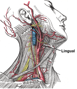
The lingual artery arises from the external carotid artery between the superior thyroid artery and facial artery.[1] It can be located easily in the tongue.

| Lingual artery | |
|---|---|
 Depiction of the neck with muscles and arteries shown. The lingual artery arises from the external carotid artery | |
 Veins of the tongue. The hypoglossal nerve has been displaced downward in this preparation (lingual artery labeled at center left). | |
| Details | |
| Source | External carotid artery |
| Branches | Sublingual artery Deep lingual artery |
| Vein | Lingual vein |
| Supplies | Genioglossus |
| Identifiers | |
| Latin | arteria lingualis |
| TA98 | A12.2.05.015 |
| TA2 | 4383 |
| FMA | 49526 |
| Anatomical terminology | |
Structure
editThe lingual artery first branches off from the external carotid artery.[1][2] It runs obliquely upward and medially to the greater horns of the hyoid bone.[1]
It then curves downward and forward, forming a loop which is crossed by the hypoglossal nerve. It then passes beneath the digastric muscle and stylohyoid muscle running horizontally forward, beneath the hyoglossus.[1][3] This takes it through the sublingual space.[4] Finally, ascending almost perpendicularly to the tongue, it turns forward on its lower surface as far as the tip of the tongue, now called the deep lingual artery (profunda linguae).
Branches
editThe lingual artery gives 4 main branches: the deep lingual artery, the sublingual artery, the suprahyoid branch, and the dorsal lingual branch.[1]
Deep lingual artery
editThe deep lingual artery (or ranine artery) is the terminal portion of the lingual artery after the sublingual artery is given off. As seen in the picture, it travels superiorly in a tortuous course along the under (ventral) surface of the tongue, below the longitudinalis inferior, and above the mucous membrane.
It lies on the lateral side of the genioglossus, the main large extrinsic tongue muscle, accompanied by the lingual nerve. However, as seen in the picture, the deep lingual artery passes inferior to the hyoglossus (the cut muscle on the bottom) while the lingual nerve (not pictured) passes superior to it (for a comparison, the hypoglossal nerve, pictured, passes superior to the hyoglossus). At the tip of the tongue, it is said to anastomose with the artery of the opposite side,[1] but this is denied by Hyrtl.[citation needed] In the mouth, these vessels are placed one on either side of the frenulum linguæ.
Sublingual artery
editThe sublingual artery arises at the anterior margin of the hyoglossus, and runs forward between the genioglossus and mylohyoid muscle to the sublingual gland.[3]
It supplies the gland and gives branches to the mylohyoideus and neighboring muscles, and to the mucous membrane of the mouth and gums.
One branch runs behind the alveolar process of the mandible in the substance of the gum to anastomose with a similar artery from the other side; another pierces the mylohyoideus and anastomoses with the submental branch of the facial artery.
Other branches
edit- The suprahyoid branch of the lingual artery runs along the upper border of the hyoid bone, supplying oxygenated blood to the muscles attached to it and joining (anastomosing) with its fellow of the opposite side.
- The dorsal lingual branches of lingual artery consist usually of two or three small branches which arise beneath the hyoglossus . They ascend medially to the back part of the dorsum of the tongue .[5] They supply the mucous membranes, the glossopalatine arch, the tonsil, soft palate, and epiglottis; anastomosing with the vessels of the opposite side.
Function
editThe lingual artery supplies the tongue.[6] It also supplies the palatine tonsils.[7]
Additional images
edit-
Lingual artery
-
Lingual artery
References
editThis article incorporates text in the public domain from page 553 of the 20th edition of Gray's Anatomy (1918)
- ^ a b c d e f Resnik, Randolph R. (2018-01-01), Resnik, Randolph R.; Misch, Carl E. (eds.), "7 - Intraoperative Complications: Bleeding", Misch's Avoiding Complications in Oral Implantology, Mosby, pp. 267–293, doi:10.1016/b978-0-323-37580-1.00007-x, ISBN 978-0-323-37580-1, retrieved 2020-11-12
- ^ Chi, T. Linda; Mirsky, David M.; Bello, Jacqueline A.; Ferson, David Z. (2013-01-01), Hagberg, Carin A. (ed.), "Chapter 2 - Airway Imaging: Principles and Practical Guide", Benumof and Hagberg's Airway Management (Third Edition), Philadelphia: W.B. Saunders, pp. 21–75.e1, doi:10.1016/b978-1-4377-2764-7.00002-6, ISBN 978-1-4377-2764-7, retrieved 2020-11-12
- ^ a b Cramer, Gregory D. (2014-01-01), Cramer, Gregory D.; Darby, Susan A. (eds.), "Chapter 5 - The Cervical Region", Clinical Anatomy of the Spine, Spinal Cord, and Ans (Third Edition), Saint Louis: Mosby, pp. 135–209, doi:10.1016/b978-0-323-07954-9.00005-0, ISBN 978-0-323-07954-9, retrieved 2020-11-12
- ^ Resnik, Randolph R.; Cillo, Joseph E. (2018-01-01), Resnik, Randolph R.; Misch, Carl E. (eds.), "8 - Intraoperative Complications: Infection", Misch's Avoiding Complications in Oral Implantology, Mosby, pp. 294–328, doi:10.1016/b978-0-323-37580-1.00008-1, ISBN 978-0-323-37580-1, retrieved 2020-11-12
- ^ Kezirian, Eric J. (2009-01-01), Eisele, David W.; Smith, Richard V. (eds.), "CHAPTER 29 - Complications of Sleep Surgery", Complications in Head and Neck Surgery (Second Edition), Philadelphia: Mosby, pp. 331–342, doi:10.1016/b978-141604220-4.50033-x, ISBN 978-1-4160-4220-4, retrieved 2020-11-12
- ^ Jacob, S. (2008-01-01), Jacob, S. (ed.), "Chapter 7 - Head and neck", Human Anatomy, Churchill Livingstone, pp. 181–225, doi:10.1016/b978-0-443-10373-5.50010-5, ISBN 978-0-443-10373-5, retrieved 2020-11-12
- ^ Witt, Martin (2019-01-01), Doty, Richard L. (ed.), "Chapter 10 - Anatomy and development of the human taste system", Handbook of Clinical Neurology, Smell and Taste, 164, Elsevier: 147–171, doi:10.1016/b978-0-444-63855-7.00010-1, ISBN 9780444638557, PMID 31604544, S2CID 204332286, retrieved 2020-11-12
External links
edit- Radiology image: Headneck:15Commo from Radiology Atlas at SUNY Downstate Medical Center (need to enable Java)
- Anatomy photo:25:14-0102 at the SUNY Downstate Medical Center
- Branches at University of Oklahoma