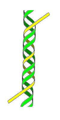
Triple-stranded DNA is a DNA structure in which three oligonucleotides wind around each other and form a triple helix. In this structure, one strand binds to a B-form DNA double helix through Hoogsteen or reversed Hoogsteen hydrogen bonds.
For example, a nucleobase T binds to a Watson–Crick base-pairing of T-A by Hoogsteen hydrogen bonds between an AxT pair (x represents a Hoogsteen base pair). An N-3 protonated cytosine, represented as C+, can also form a base-triplet with a C-G pair through the Hoogsteen base-pairing of an GxC+. Thus, the triple-helical DNAs using these Hoogsteen pairings consist of two homopyrimidines and one homopurine, and the homopyrimidine third strand is parallel to the homopurine strand.
A homopurine third strand can also bind to a homopurine-homopyrimidine duplex using reversed Hoogsteen patterns. In this triplex, a nucleobase A binds to a T-A base pair and a G to a C-G pair. Since the nucleobases on the third strand have to be reversed, the homopurine third strand is antiparallel to the homopurine strand of the original duplex.
History
editTriple-stranded DNA was a common hypothesis in the 1950s when scientists were struggling to discover DNA's true structural form. Watson and Crick (who later won the Nobel Prize for their double-helix model) originally considered a triple-helix model, as did Pauling and Corey, who published a proposal for their triple-helix model in 1953, as well as fellow scientist Fraser. However, Watson and Crick soon identified several problems with these models:
- Negatively charged phosphates near the axis repel each other, leaving the question of how the three-chain structure stays together.
- In a triple-helix model (specifically Pauling and Corey's model), some of the van der Waals distances appear to be too small.
Fraser's model differed from Pauling and Corey's in that in his model the phosphates are on the outside and the bases are on the inside, linked together by hydrogen bonds. However, Watson and Crick found Fraser's model to be too ill-defined to comment specifically on its inadequacies.
Triple-stranded DNA was also described in 1957, when it was thought to occur in only one in vivo biological process: as an intermediate product during the action of the E. coli recombination enzyme RecA. Its role in that process is not understood.
Using nucleic acid segments that bind to the DNA duplexes to form triple strands as a way of regulating gene expression is under separate investigation by biotechnology companies and Yale University.
Function
editTriple-stranded DNA is implicated in the regulation of several genes. Once, the c-myc gene was extensively mutated to examine the role that triplex DNA structure, versus linear sequence, plays in gene regulation. A c-myc promoter element, termed the nuclease-sensitive element or NSE, can form tandem intramolecular triplexes of the H-DNA type and has a repeating sequence motif (ACCCTCCCC)4. The NSE when mutated was examined for transcriptional activity and for intra- and intermolecular triplex forming ability. The transcriptional activity of mutant NSEs can be predicted by the element's ability to form H-DNA and not by repeat number, position, or the number of mutant base pairs. DNA may therefore be a dynamic participant in the transcription of the c-myc gene.[1]
A DNA triplex is formed when pyrimidine or purine bases occupy the major groove of the DNA double Helix forming Hoogsteen pairs with purines of the Watson-Crick basepairs. Intermolecular triplexes are formed between triplex-forming oligonucleotides (TFO) and target sequences on duplex DNA. Intramolecular triplexes are the major elements of H-DNAs, unusual DNA structures that are formed in homopurine-homopyrimidine regions of supercoiled DNAs.Triplex forming oligonucleotides (TFOs) consist of sequences of DNA that are rich in polypurine and polypyrimidine sequences. These regions have a much larger chance of forming triplex DNA. Triplexes have been shown to function in the process of transcription, as a result, triplexes can alter or impair the transcription of certain genes. A large percentage of non-B-DNA structures are associated with genes that correspond to the brain and neurological system. Triplexes also have higher frequencies of double strand breaks, which can cause genomic rearrangements like insertions, translocations, inversions, and gross deletions. This genomic instability is a result of the triplex DNA supercoiling to overcome its highly unfavorable thermodynamic constraints. This genomic instability of non-B-DNA is the cause for many neurological diseases, like Fredrick’s Ataxia, which is a recessive autosomal trait that results in the progressive degeneration of the nervous system and spinal cord, impairing the movement of the limbs. Fredrick’s Ataxia is a direct result of impaired transcription by intramolecular triplex DNA.
TFOs are promising gene-drugs that can be used in an anti-gene strategy. They attempt to modulate gene activity in vivo. Chemical modifications of TFO are known. In peptide nucleic acid (PNA), the sugar-phosphate backbone is replaced with a protein-like backbone. PNAs form P-loops while interacting with duplex DNA, forming triplex with one DNA strand displacing the other. Very unusual recombination or parallel triplexes, or R-DNA, have been assumed to form under RecA protein in the course of homologous recombination.[2]
References
edit- ^ Firulli, A.B.; Maibenco, D.C.; Kinniburgh, A.J. (1994). "Triplex Forming Ability of a c-myc Promoter Element Predicts Promoter Strength". Archives of Biochemistry and Biophysics. 310 (1): 236–42. doi:10.1006/abbi.1994.1162. PMID 8161210.
- ^ Frank-Kamenetskii, M. D.; Mirkin, S. M. (1995-01-01). "Triplex DNA structures". Annual Review of Biochemistry. 64: 65–95. doi:10.1146/annurev.bi.64.070195.000433. ISSN 0066-4154. PMID 7574496.
Sources
editThis article includes a list of general references, but it lacks sufficient corresponding inline citations. (May 2017) |
- Rich, Alexander (1993). "DNA comes in many forms". Gene. 135 (1–2): 99–109. doi:10.1016/0378-1119(93)90054-7. PMID 8276285.
- Soyfer, Valery N.; Potaman, Vladimir N. (1995). Triple-Helical Nucleic Acids. New York: Springer. ISBN 978-0-387-94495-1.
- Mills, Martin; Arimondo, Paola B.; Lacroix, Laurent; Garestier, Thérèse; Hélène, Claude; Klump, Horst; Mergny, Jean-Louis (1999). "Energetics of strand-displacement reactions in triple helices: A spectroscopic study". Journal of Molecular Biology. 291 (5): 1035–54. doi:10.1006/jmbi.1999.3014. PMID 10518941.
- Watson, J. D.; Crick, F. H. C. (1953). "Molecular Structure of Nucleic Acids: A Structure for Deoxyribose Nucleic Acid". Nature. 171 (4356): 737–8. Bibcode:1953Natur.171..737W. doi:10.1038/171737a0. PMID 13054692.