This article needs more reliable medical references for verification or relies too heavily on primary sources. (March 2023) |  |
Clinical pathology is a medical specialty that is concerned with the diagnosis of disease based on the laboratory analysis of bodily fluids, such as blood, urine, and tissue homogenates or extracts using the tools of chemistry, microbiology, hematology, molecular pathology, and Immunohaematology. This specialty requires a medical residency.


Clinical pathology is a term used in the US, UK, Ireland, many Commonwealth countries, Portugal, Brazil, Italy, Japan, and Peru; countries using the equivalent in the home language of "laboratory medicine" include Austria, Germany, Romania, Poland and other Eastern European countries; other terms are "clinical analysis" (Spain) and "clinical/medical biology (France, Belgium, Netherlands, North and West Africa).[1]
Licensing and subspecialities
editThe American Board of Pathology certifies clinical pathologists, and recognizes the following secondary specialties of clinical pathology:
- Chemical pathology, also called clinical chemistry
- Hematopathology
- Blood banking - Transfusion medicine
- Clinical microbiology
- Cytogenetics
- Molecular genetics pathology.
In some countries other sub specialities fall under certified Clinical Biologists responsibility:[2]
Organization
editClinical pathologists are often medical doctors. In some countries in South-America, Europe, Africa or Asia, this specialty can be practiced by non-physicians, such as Ph.D. or Pharm.D. after a variable number of years of residency.
In United States of America
editClinical pathologists work in close collaboration with clinical scientists (clinical biochemists, clinical microbiologists, etc.), medical technologists, hospital administrators, and referring physicians to ensure the accuracy and optimal utilization of laboratory testing.
Clinical pathology is one of the two major divisions of pathology, the other being anatomical pathology. Often, pathologists practice both anatomical and clinical pathology, a combination sometimes known as general pathology. Similar specialties exist in veterinary pathology.
Clinical pathology is itself divided into subspecialties, the main ones being clinical chemistry, clinical hematology/blood banking, hematopathology and clinical microbiology and emerging subspecialties such as molecular diagnostics and proteomics. Many areas of clinical pathology overlap with anatomic pathology. Both can serve as medical directors of CLIA certified laboratories. Under the CLIA law, only the US Department of Health and Human Services approved Board Certified Ph.D., DSc, or MD and DO can perform the duties of a Medical or Clinical Laboratory Director. This overlap includes immunoassays, flow cytometry, microbiology and cytogenetics and any assay done on tissue. Overlap between anatomic and clinical pathology is expanding to molecular diagnostics and proteomics as we move towards making the best use of new technologies for personalized medicine.[3]
Clinical pathologists may assist physicians in interpreting complex tests such as platelet aggregometry, hemoglobin or serum protein electrophoresis, or coagulation profiles. If interfering substances are suspected, they may recommend alternate test methods. For example, hemolysis, icterus, lipemia, or heterophile antibodies may confound results obtained by traditional methods such as ion-selective electrodes, enzymatic assays or immunoassays. Alternate methods such as blood gas analysers, point-of-care testing or mass spectrometry may help resolve the clinical question.
In Europe
editRecently, EFLM has chosen the name of "Specialists in Laboratory Medicine" to define all European Clinical pathologists, regardless of their training (M.D., Ph.D. or Pharm.D.).[4]
In France, Clinical Pathology is called Medical Biology ("Biologie médicale") and is practiced by both M.D.s and Pharm.D.s. The residency lasts four years. Specialists in this discipline are called "Biologiste médical" which literally translates as Clinical Biologist rather than "Clinical pathologist".[5]
Tools
editMicroscopes, analyzers, strips, centrifuges
Macroscopic examination
editVisual examination of the specimen may provide information to the pathologist or the physician. For example, fluid drained from an abscess may appear cloudy, or cerebrospinal fluid obtained by lumbar puncture may exhibit xanthochromia, suggesting a bleed has occurred. Laboratory technologists may provide qualitative descriptions accordingly.
Microscopical examination
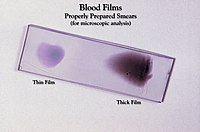
editMicroscopic analysis is an important activity of the pathologist and the laboratory technologist. They have many different stains at their disposal (GRAM, MGG, Grocott, Ziehl–Neelsen, etc.). Immunofluorescence, cytochemistry, the immunocytochemistry, and FISH are also used in order make a correct diagnosis.
Pathologists may review samples such as pleural, peritoneal, synovial, or pericardial fluids to characterize them as "normal", tumoral, inflammatory, or even infectious. Microscopic examination can also determine the causal infectious agent – often a bacterium, mould, yeast, parasite, or (rarely) virus.
Laboratory Analysers
editAutomated analysers, by the association of robotics and spectrophotometry, have allowed these last decades better reproducibility of the results, in particular in medical biochemistry and hematology.[6]
Efficiency and productivity can be enhanced by automating the pre-analytical processing, including barcode reading, sorting, centrifuging, and aliquoting specimens.
The analysers must undergo daily controls prior to performing patient testing. Analysers must also undergo daily, weekly and monthly maintenance. Quality management involves reviewing quality control trends to detect emerging problems in instrument calibration, correlating results between instruments that perform similar testing, and running standardized samples to prove linearity and precision.
Some laboratory processes involve automated analysis combined with manual review by technologists. For example, when hematology analysers flag samples as abnormal, automated white blood cell differential counts may be superseded by manual differential counts using stained slides read at the microscope or scanned by digital imaging software. Laboratory technologists may flag abnormal samples for pathologist review. The pathologist may recommend additional testing, such as flow cytometry to identify lymphoma or leukemia cells, or cytology to characterize solid tumor cells.
Cultures
editSamples undergoing examination for pathogens, primarily in medical microbiology, may be incubated with culture media. Those allow, for example, the description of one or several infectious agents responsible of the clinical signs.
Values known as "normal" or reference values
editDetailed article: Reference range.
See also
editNotes and references
edit- ^ "Textes Généraux, Ministère de la Santé et des Sports". Journal Officiel de la République Française. Décrets, arrêtés, circulaires (Texte 15 sur 54). 20 June 2010. Retrieved 4 December 2019. Note: This document does not cover all countries listed.
- ^ "Bulletin officiel du n°32 du 4 septembre 2003 - MENS0301444A". www.education.gouv.fr. Retrieved 2023-02-21.
- ^ Description of Pathology in USA
- ^ Zerah Simone, Murray Janet, Rita Horvath Andrea (2012). "EFLM Position Statement – Our profession now has a European name: Specialist in Laboratory Medicine". Biochemia Medica. 22 (3): 272–273. doi:10.11613/BM.2012.029. PMC 3900053. PMID 23092058.
{{cite journal}}: CS1 maint: multiple names: authors list (link) - ^ Reglementation for French Residency in Clinical Pathology Archived 2008-02-28 at the Wayback Machine
- ^ More D, Khan N, Tekade RK, Sengupta P. An Update on Current Trend in Sample Preparation Automation in Bioanalysis: strategies, Challenges and Future Direction. Crit Rev Anal Chem. 2024 Jul 1:1-25. doi: 10.1080/10408347.2024.2362707. Epub ahead of print. PMID: 38949910.