This article needs additional citations for verification. (April 2018) |
| Portal vein thrombosis | |
|---|---|
 | |
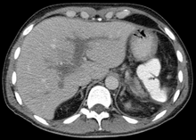
| Portal vein thrombosis seen with computed tomography. |
Portal vein thrombosis is a condition that occurs when a blood clot occurs in the hepatic portal vein, which can lead to increased pressure in the portal vein system and reduced blood supply to the liver.
Signs and symptoms
editPortal vein thrombosis causes upper abdominal pain, possibly accompanied by fever, nausea, and enlarged liver and/or spleen; the abdomen may be filled with fluid (ascites). However, it can also develop without causing symptoms, leading to portal hypertension before it is diagnosed.[1] Other symptoms can develop based on the cause. For example, if portal vein thrombosis develops due to liver cirrhosis, bleeding or other signs of liver disease may be present. If portal vein thrombosis develops due to pylephlebitis, signs of infection such as fever, chills, night sweats may be present.
Causes
editSlowed blood flow due to underlying cirrhosis or congestive heart failure is often implicated. Thrombophilia (including inherited conditions such as factor V Leiden deficiency, protein C or S deficiency, or antiphospholipid antibody syndrome) is another common cause. Non-inherited tendency for thrombosis may be due to underlying myeloproliferative disorders, oral contraceptive use, or pregnancy. Alternatively, the portal vein may be injured as a result of pancreatitis, diverticulitis, cholangiocarcinoma, or abdominal surgery/trauma. PVT is also a known complication of surgical removal of the spleen.[2] During the last several years, myeloproliferative neoplasms (MPNs) have emerged as a leading systemic cause of splanchnic vein thromboses (includes PVT).
Mechanism
editThe main portal vein is formed by the union of the splenic vein and superior mesenteric vein (SMV). It is responsible for approximately three-fourths of the liver’s blood flow, transported from much of the gastrointestinal system as well as the pancreas, gallbladder, and spleen. Cirrhosis alters bleeding pathways thus patients are simultaneously at risk of uncontrolled bleeding and forming clots.
Diagnosis
editThe diagnosis of portal vein thrombosis is usually made with imaging, of which Doppler ultrasound is the least invasive. Determination of severity may be derived via computed tomography (CT) with contrast, magnetic resonance imaging (MRI), or MR angiography (MRA). Those with chronic PVT may undergo upper endoscopy (esophagogastroduodenoscopy, EGD) to evaluate the presence of concurrent dilated veins (varices) in the stomach or esophagus. D-dimer levels in the blood may be elevated as a result of fibrin breakdown.
Treatment
editUnless there are underlying reasons why it would be harmful, anticoagulation (low molecular weight heparin, followed by warfarin) is often initiated and maintained in patients who do not have cirrhosis. Anticoagulation for patients with cirrhosis who experience portal vein thrombosis is usually not advised unless they have chronic PVT 1) with thrombophilia, 2) with clot burden in the mesenteric veins, or 3) inadequate blood supply to the bowels. In more severe instances, shunts or a liver transplant may be considered.
See also
editReferences
edit- ^ Hall, TC; Garcea, G; Metcalfe, M; Bilku, D; Dennison, AR (November 2011). "Management of acute non-cirrhotic and non-malignant portal vein thrombosis: a systematic review". World Journal of Surgery. 35 (11): 2510–20. doi:10.1007/s00268-011-1198-0. PMID 21882035.
- ^ Ali Cadili, Chris de Gara, "Complications of Splenectomy", The American Journal of Medicine, 2008, pp 371-375.
External links
editThis is a user sandbox of Mizrebel83. You can use it for testing or practicing edits. This is not the sandbox where you should draft your assigned article for a dashboard.wikiedu.org course. To find the right sandbox for your assignment, visit your Dashboard course page and follow the Sandbox Draft link for your assigned article in the My Articles section. |